
| Professional Committee of Rock and Mineral Testing Technology of the Geological Society of China, National Geological Experiment and Testing Center | Host |
| Citation: |
SUN Chengyang, LU Taijin, SONG Zhonghua, HE Mingyue, DENG Yi. Analysis of Abnormal Birefringence and Graphite Inclusions in Zimbabwean Diamonds[J]. Rock and Mineral Analysis, 2022, 41(2): 199-210. doi: 10.15898/j.cnki.11-2131/td.202111050165

|
Analysis of Abnormal Birefringence and Graphite Inclusions in Zimbabwean Diamonds
-
Abstract
BACKGROUND The Marange diamond deposit in Zimbabwe is characterized by producing mixed-habit (octahedral and cuboid) diamonds. Graphite inclusions in these diamonds only exist in cuboid sectors. The morphological and distributional characteristics of graphite inclusions and the abnormal birefringence and strain characteristics of diamonds can reflect the geological process experienced by diamonds from the beginning of crystallization to being transported to the Earth's surface. Therefore, the study of diamonds and graphite inclusions in Zimbabwe can provide comparative data for diamonds from other deposits. Besides, due to the peculiarity of growth habits, detailed analysis would be of great value to help understand the behavioral differences of diamonds with different growth habits in geological processes.
OBJECTIVES To determine if graphite inclusions in Zimbabwean diamonds are syngenetic or epigenetic, and to reveal the relationship between graphite inclusions and the infrared absorption spectrum, Raman scattering spectrum as well as birefringence and strain characteristics of diamonds.
METHODS The growth structure and growth sectors of Zimbabwean diamonds were observed by DiamondViewTM image system. The morphological and distributional characteristics of graphite inclusions and abnormal birefringence in diamonds were analyzed by scanning electron microscopy (SEM) and polarized light microscopy. Analysis of distribution and relative concentration of impurity elements in different growth sectors was conducted by infrared spectroscopy. Strain characteristics of diamonds in different growth sectors were analyzed by Raman spectroscopy and projection diagram of corresponding results.
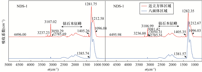
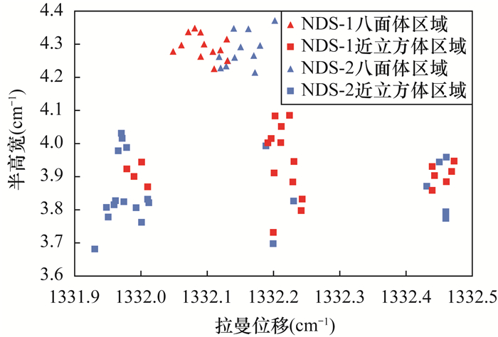
RESULTS Graphite inclusions in cuboid sectors of Zimbabwean diamonds were syngenetic-epigenetic inclusions located in directional elliptical cracks. According to infrared spectra of different growth sectors, cuboid sectors showed stronger infrared absorption related to elemental hydrogen, while octahedral sectors showed stronger absorption related to elemental nitrogen. This enrichment of different impurity elements leading to abnormal birefringence was mainly related to cracks and different growth sectors in diamond. The Raman shift of LO=TO band in octahedral sectors was 1332.05-1332.20cm-1, the FWHM was 4.21-4.37cm-1, which corresponded to stress of 0.06-0.27GPa. The Raman shift of LO=TO band in cuboid sectors was 1331.93-1332.47cm-1, the FWHM was 3.67-4.08cm-1, which corresponded to stress of 0.01-0.64GPa. In general, the residual stress and strain were greater in cuboid sectors.
CONCLUSIONS The determination of the orientation of graphite inclusions in mixed-habit diamonds in Zimbabwe, provides new evidence to prove their syngenetic-epigenetic nature, and reveal the difference in the strain characteristics of diamonds in the two growth regions. This research is helpful for understanding the formation environment of diamonds in Zimbabwe and of different diamonds. The differences in physicochemical properties are of great significance.
-

-
References
[1] Zedgenizov D A, Kagi H, Shatsky V S, et al. Carbonatitic melts in cuboid diamonds from Udachnaya kimberlite pipe (Yakutia): Evidence from vibrational spectroscopy[J]. Mineralogical Magazine, 2004, 68(1): 61-73. doi: 10.1180/0026461046810171 [2] Skuzovatov S Yu, Zedgenizov D A, Shatsky V S, et al. Composition of cloudy microinclusions in octahedral diamonds from the Internatsional'naya kimberlite pipe (Yakutia)[J]. Russian Geology and Geophysics, 2011, 52(1): 85-96. doi: 10.1016/j.rgg.2010.12.007 [3] Lang A R. Causes of birefringence in diamond[J]. Nature, 1967, 213(5073): 248-251. doi: 10.1038/213248a0 [4] Barron L M, Mernagh T P, Barron B J. Using strain birefringence in diamond to estimate the remnant pressure on an inclusion[J]. Australian Journal of Earth Sciences, 2008, 55(2): 159-165. doi: 10.1080/08120090701689332 [5] Rosenfeld J L, Chase A B. Pressure and temperature of crystallization from elastic effects around solid inclusions in minerals?[J]. American Journal of Science, 1961, 259(7): 519-541. doi: 10.2475/ajs.259.7.519 [6] Howell D, Wood I G, Dobson D P, et al. Quantifying strain birefringence halos around inclusions in diamond[J]. Contributions to Mineralogy and Petrology, 2010, 160(5): 705-717. doi: 10.1007/s00410-010-0503-5 [7] 陆太进, 陈华, 张健, 等. 津巴布韦金刚石独特的形态及其"指纹"特征的意义[J]. 地质通报, 2011, 31(10): 25-32. Lu T J, Chen H, Zhang J, et al. Unique morphology of Zimbabwe diamond and its 'fingerprint' characteristic significance[J]. Geological Bulletin of China, 2011, 30(10): 25-32. [8] Rondeau B, Fritsch E, Guiraud M, et al. Three historical 'asteriated' hydrogen-rich diamonds: Growth history and sector-dependent impurity incorporation[J]. Diamond and Related Materials, 2004, 13(9): 1658-1673. doi: 10.1016/j.diamond.2004.02.002 [9] Howell D, Griffin W L, Piazolo S, et al. A spectroscopic and carbon-isotope study of mixed-habit diamonds: Impurity characteristics and growth environment[J]. American Mineralogist, 2013, 98(1): 66-77. doi: 10.2138/am.2013.4179 [10] Smit K V, Shirey S B, Stern R A, et al. Diamond growth from C-H-N-O recycled fluids in the lithosphere: Evidence from CH4 micro-inclusions and δ 13C- δ 15N-N content in Marange mixed-habit diamonds[J]. Lithos, 2016, 265: 68-81. doi: 10.1016/j.lithos.2016.03.015 [11] Smit K V, Myagkaya E, Persaud S, et al. Black diamonds from Marange (Zimbabwe): A result of natural irradiation and graphite inclusions[J]. Gems and Gemology, 2018, 54(2): 132-148. doi: 10.5741/GEMS.54.2.132 [12] Pal'Yanov Y N, Sokol A G, Sobolev N V. Experimental modeling of mantle diamond-forming processes[J]. Russian Geology and Geophisics, 2005, 46(12): 1290-1303. [13] Sokol A G, Pal'Yanov Y N. Diamond formation in the system MgO-SiO2-H2O-C at 7.5GPa and 1600℃[J]. Contributions to Mineralogy and Petrology, 2008, 155(1): 33-43. [14] Zedgenizov D A, Ragozin A L, Shatsky V S, et al. Fibrous diamonds from the placers of the northeastern Siberian Platform: Carbonate and silicate crystallization media[J]. Russian Geology and Geophysics, 2011, 52(11): 1298-1309. doi: 10.1016/j.rgg.2011.10.003 [15] Izraeli E S, Harris J W, Navon O. Brine inclusions in diamonds: A new upper mantle fluid[J]. Earth and Planetary Science Letters, 2001, 187(3-4): 323-332. doi: 10.1016/S0012-821X(01)00291-6 [16] Harris J W. Black material on mineral inclusions and in internal fracture planes in diamond[J]. Contributions to Mineralogy and Petrology, 1972, 35(1): 22-33. doi: 10.1007/BF00397374 [17] Nechaev D V, Khokhryakov A F. Formation of metastable graphite inclusions during diamond crystallization in model systems[J]. Geology of Ore Deposits, 2014, 56(2): 139-146. doi: 10.1134/S1075701514020044 [18] Khokhryakov A F, Nechaev D V, Sokol A G, et al. Formation of various types of graphite inclusions in diamond: Experimental data[J]. Lithos, 2009, 112: 683-689. doi: 10.1016/j.lithos.2009.05.010 [19] Nechaev D V, Khokhryakov A F. Formation of epigenetic graphite inclusions in diamond crystals: Experimental data[J]. Russian Geology and Geophysics, 2013, 54(4): 399-405. doi: 10.1016/j.rgg.2013.03.003 [20] 唐诗, 苏隽, 陆太进, 等. 化学气相沉积法再生钻石的实验室检测特征研究[J]. 岩矿测试, 2019, 38(1): 62-70. Tang S, Su J, Lu T J, et al. Research on laboratory testing features of chemical vapor deposition in overgrowth diamonds[J]. Rock and Mineral Analysis, 2019, 38(1): 62-70. [21] Rakovan J, Gaillou E, Post J E, et al. Optically sector-zoned (star) diamonds from Zimbabwe[J]. Rock and Minerals, 2014, 89(2): 173-178. doi: 10.1080/00357529.2014.842844 [22] Nasdala L, Brenker F E, Glinnemann J, et al. Spectroscopic 2D-tomography: Residual pressure and strain around mineral inclusions in diamonds[J]. European Journal of Mineralogy, 2003, 15(6): 931-935. [23] Howell D. Strain-induced birefringence in natural diamond: A review[J]. European Journal of Mineralogy, 2012, 24(4): 575-585. doi: 10.1127/0935-1221/2012/0024-2205 [24] Gogotsi Y G, Kailer A, Nickel K G. Transformation of diamond to graphite[J]. Nature, 1999, 401(6754): 663-664. doi: 10.1038/44323 [25] Day H W. A revised diamond-graphite transition curve[J]. American Mineralogist, 2012, 97(1): 2-62. [26] 杨志军, 彭明生, 谢先德, 等. 金刚石的微区显微红外光谱分析及其意义[J]. 岩矿测试, 2002, 21(3): 161-165. doi: 10.3969/j.issn.0254-5357.2002.03.001 Yang Z J, Peng M S, Xie X D, et al. Micro area analysis of diamond by micro-infrared spectrometry and its significance[J]. Rock and Mineral Analysis, 2002, 21(3): 161-165. doi: 10.3969/j.issn.0254-5357.2002.03.001 [27] Zaitsev A M. Optical properties of diamond: A data handbook[M]. Springer Science and Business Media, 2013: 52-57. [28] Goss J P, Briddon P R, Hill V, et al. Identification of the structure of the 3107cm-1 H-related defect in diamond[J]. Journal of Physics: Condensed Matter, 2014, 26(14): 145801. doi: 10.1088/0953-8984/26/14/145801 [29] Salustro S, Gentile F S, D'Arco P, et al. Hydrogen atoms in the diamond vacancy defect. A quantum mechanical vibrational analysis[J]. Carbon, 2018, 129(1): 349-356. [30] 宋中华, 陆太进, 苏隽, 等. 利用吸收和发光光谱技术分析高温高压天然富氢钻石的鉴定特征[J]. 岩矿测试, 2018, 37(1): 64-69. Song Z H, Lu T J, Su J, et al. Identification of HPHT-treated hydrogen-rich diamonds by optical absorption and photo luminescence spectroscopy techniques[J]. Rock and Mineral Analysis, 2018, 37(1): 64-69. [31] Davies G, Collins A T, Spear P. Sharp infra-red absorption lines in diamond[J]. Solid State Communications, 1984, 49(5): 433-436. doi: 10.1016/0038-1098(84)90657-4 [32] Benedetti L R, Nguyen J H, Caldwell W A, et al. Dissociation of CH4 at high pressures and temperatures: Diamond formation in giant planet interiors?[J]. Science, 1999, 286(5437): 100-102. doi: 10.1126/science.286.5437.100 [33] Peaker C V, Goss J P, Briddon P R, et al. Di-nitrogen-vacancy-hydrogen defects in diamond: A computational study[J]. Physica Status Solid A, 2015, 212(11): 2616-2620. doi: 10.1002/pssa.201532216 [34] Gu T, Wang W. Optical defects in milky type aB diamonds [J]. Diamond and Related Materials, 2018, 89: 322-329. doi: 10.1016/j.diamond.2018.09.010 [35] Clackson S G, Moore M, Walmsley J, et al. The relationship between platelet size and the frequency of the B'infrared absorption peak in type Ⅰa diamond[J]. Philosophical Magazine B, 1990, 62(2): 115-128. doi: 10.1080/13642819008226980 [36] Collinss A T, Kanda H, Burns R C. The segregation of nickel-related optical centres in the octahedral growth sectors of synthetic diamond[J]. Philosophical Magazine B, 1990, 61(5): 797-810. doi: 10.1080/13642819008207562 [37] Burns R C, Cvetkovic V, Dodge C N, et al. Growth-sector dependence of optical features in large synthetic diamonds[J]. Journal of Crystal Growth, 1990, 104(2): 257-279. doi: 10.1016/0022-0248(90)90126-6 [38] Boyd S R, Pillinger C T, Milledge H J, et al. Fractionation of nitrogen isotopes in a synthetic diamond of mixed crystal habit[J]. Nature, 1988, 331(6157): 604-607. doi: 10.1038/331604a0 [39] Boyd S R, Kiflawi I, Woods G S. The relationship between infrared absorption and the A defect concentration in diamond[J]. Philosophical Magazine B, 1994, 69(6): 1149-1153. doi: 10.1080/01418639408240185 [40] Boyd S R, Kiflawi I, Woods G S. Infrared absorption by the B nitrogen aggregate in diamond[J]. Philosophical Magazine B, 1995, 72(3): 351-361. doi: 10.1080/13642819508239089 [41] Kiflawi I, Kanda H, Fisher D, et al. The aggregation of nitrogen and the formation of A centres in diamonds[J]. Diamond and Related Materials, 1997, 6(11): 1643-1649. doi: 10.1016/S0925-9635(97)00207-0 [42] Fisher D, Lawson S C. The effect of nickel and cobalt on the aggregation of nitrogen in diamond[J]. Diamond and Related Materials, 1998, 7(2): 299-304. [43] Lang A R, Bulanova G P, Fisher D, et al. Defects in a mixed-habit Yakutian diamond: Studies by optical and cathodoluminescence microscopy, infrared absorption, Raman scattering and photoluminescence spectroscopy[J]. Journal of Crystal Growth, 2007, 309(2): 170-180. doi: 10.1016/j.jcrysgro.2007.09.022 [44] Grimsditch M H, Anastassakis E, Cardona M. Piezo-birefringence in diamond[J]. Physical Review B, 1979, 19(6): 3240-3243. doi: 10.1103/PhysRevB.19.3240 [45] Higashida K, Tanaka M, Matsunaga E, et al. Crack tip stress fields revealed by infrared photoelasticity in silicon crystals[J]. Materials Science and Engineering A, 2004, 387: 377-380. [46] Welbourn C M, Rooney M, Evans D. A study of dia-monds of cube and cube-related shape from the Jwaneng mine[J]. Journal of Crystal Growth, 1989, 94(1): 229-252. doi: 10.1016/0022-0248(89)90622-2 [47] Lawn B R, Komatsu H. The nature of deformation around pressure cracks on diamond[J]. Philosophical Magazine, 1966, 14(130): 689-699. doi: 10.1080/14786436608211965 [48] Nasdala L, Hofmeister W, Harris J W, et al. Growth zoning and strain patterns inside diamond crystals as revealed by Raman maps[J]. American Mineralogist, 2005, 90(4): 745-748. doi: 10.2138/am.2005.1690 [49] Nemanich R J, Solin S A. First-and second-order Raman scattering from finite-size crystals of graphite[J]. Physical Review B, 1979, 20(2): 392. doi: 10.1103/PhysRevB.20.392 [50] 马瑛, 王琦, 丘志力, 等. 湖南砂矿金刚石中石墨包裹体拉曼光谱原位测定: 形成条件及成因指示[J]. 光谱学与光谱分析, 2018, 38(6): 1753-1757. Ma Y, Wang Q, Qiu Z L, et al. In-situ Raman spectroscopy testing and genesis of graphite inclusions in alluvial diamonds from Hunan[J]. Spectroscopy and Spectral Analysis, 2018, 38(6): 1753-1757. [51] Zerda T W, Xu W, Zerda A, et al. High pressure Raman and neutron scattering study on structure of carbon black particles[J]. Carbon, 2000, 38(3): 355-361. doi: 10.1016/S0008-6223(99)00111-6 [52] Howell D, Wood I G, Nestola F, et al. Inclusions under remnant pressure in diamond: A multi-technique approach[J]. European Journal of Mineralogy, 2012, 24(4): 563-573. doi: 10.1127/0935-1221/2012/0024-2183 [53] Grimsditch M H, Anastassakis E, Cardona M. Effect of uniaxial stress on the zone-center optical phonon of diamond[J]. Physical Review B, 1978, 18(2): 901. doi: 10.1103/PhysRevB.18.901 [54] Nachal'Naya T A, Andreyev V D, Gabrusenok E V. Shift of the frequency and Stokes-anti-Stokes ratio of Raman spectra from diamond powders[J]. Diamond and Related Materials, 1994, 3(11-12): 1325-1328. doi: 10.1016/0925-9635(94)90146-5 [55] Surovtsev N V, Kupriyanov I N. Effect of nitrogen impur-ities on the Raman line width in diamond, revisited[J]. Crystals, 2017, 7(8): 239. doi: 10.3390/cryst7080239 [56] Harris J W, Vance E R. Induced graphitisation around crystalline inclusions in diamond[J]. Contributions to Mineralogy and Petrology, 1972, 35(3): 227-234. doi: 10.1007/BF00371217 -
Access History
Figures(9)

Export File
Citation
SUN Chengyang, LU Taijin, SONG Zhonghua, HE Mingyue, DENG Yi. Analysis of Abnormal Birefringence and Graphite Inclusions in Zimbabwean Diamonds[J]. Rock and Mineral Analysis, 2022, 41(2): 199-210. doi: 10.15898/j.cnki.11-2131/td.202111050165
Format
Content
 DownLoad:
-
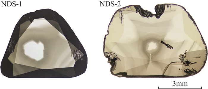
Figure 1.
Two natural diamond sections with mixed-habit from Marange, Zimbabwe
-
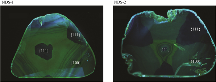
Figure 2.
Fluorescence images of diamond section samples excited by ultra-violet light of DiamondViewTM
-
Figure 3.
Distribution characteristics of inclusions in Zimbabwean diamond sections
-
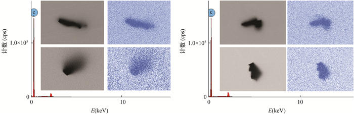
Figure 4.
Morphological characteristics of graphite inclusions in Zimbabwean diamond sections under SEM and EDX mappings of corresponding regions
-
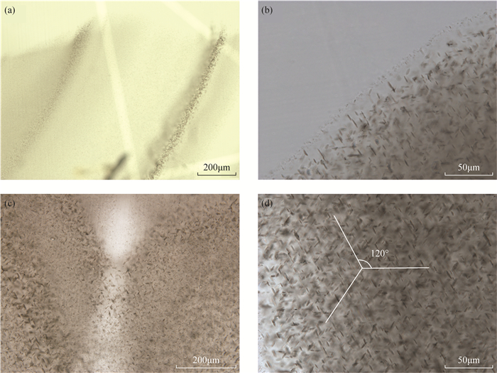
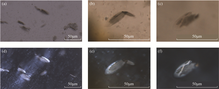
Figure 5.
Microphotographs of graphite inclusions and surrounding elliptical cracks in Zimbabwean diamonds
-
Figure 6.
Infrared absorption spectra of different growth sectors in mixed-habit diamonds from Marange, Zimbabwe
-
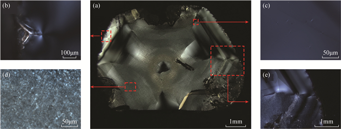
Figure 7.
Abnormal birefringence in sample NDS-2
-
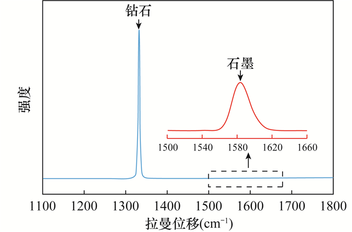
Figure 8.
Raman scattering spectrum of cuboid sectors of diamond section samples from Marange, Zimbabwe
-
Figure 9.
Projection diagram based on Raman shift and FWHM of LO=TO band of diamond sections from Marange, Zimbabwe