
| Professional Committee of Rock and Mineral Testing Technology of the Geological Society of China, National Geological Experiment and Testing Center | Host |

| Citation: |
HE Jia-le, GONG Ting-ting, PAN Zhong-xi, DU Gu. Raman Imaging Analysis Method of Fine Minerals in Rock Ore[J]. Rock and Mineral Analysis, 2021, 40(4): 491-503. doi: 10.15898/j.cnki.11-2131/td.202103080036

|
Raman Imaging Analysis Method of Fine Minerals in Rock Ore
-
Abstract
BACKGROUND Mineral identification is the basis of all types of geological work, and its appraisal level and quality directly affect the depth and degree of research of a study. Conventional identification methods are significantly influenced by experience level, optical microscope resolution, and other factors. It is difficult to accurately identify fine rare minerals and clay minerals that need to be studied. Additionally, most of the technical methods relying on high-precision large-scale instruments have special requirements for sample preparation, which is not conducive to the reuse of the samples. It is also inconvenient to explore and observe specific fine transparent minerals under high multiple reflected lights, such as scanning electron microscopy and electron microprobe.
OBJECTIVES To develop a more rapid and accurate method for identifying fine minerals.
METHODS The laser Raman high-resolution large-area fast imaging method (StreamLineHR) was applied to the whole-area large-area scanning spectrum of two standard rock slices.
RESULTS The transparent minerals were identified as alkali feldspar, plagioclase, quartz, amphibole, biotite, calcite, sphene, apatite, zircon, and epidote. The opaque mineral was identified as magnetite. Some of the minerals were closely associated (e.g., quartz and feldspar as well as sphene and hornblende), and some minerals showed secondary alterations (e.g., feldspar was transformed to calcite). Based on the content statistics, the two thin sections were named fine-grained amphibolite monzonite and fine-grained biotite plagioclase amphibolite.
CONCLUSIONS Experimental results showed that this method was more accurate than the conventional methods used for the identification of fine minerals with very low content. However, the interference caused by the fluorescence effect, similarity in peak positions of similar minerals (feldspar, amphibole), and shift of the peak position of altered minerals during mineral identification and spectral fitting were solved by combining the optical characteristics under the mineral objective lens when necessary. In addition, the smaller the setting of the surface sweep step size, the more accurate the analysis, and the time cost correspondingly increased. This method realized the rapid identification of fine minerals over a large range, which was convenient, intuitive, and accurate. It compensated for the shortcomings of conventional rock and ore identification and other technical methods and expanded the application scope of Raman spectroscopy in geological studies.
-

-
References
[1] 贾福东, 张长青, 化志新, 等. 云南麻花坪钨铍矿床蓝柱石的鉴定特征及成分与成因分析[J]. 光谱学与光谱分析, 2020, 40(10): 3185-3192. Jia F D, Zhang C Q, Hua Z X, et al. Identification characteristics, composition and genesis of euclase in Mahuaping tungsten-beryllium polymetallic deposit in Yunnan Province, southwest China[J]. Spectroscopy and Spectral Analysis, 2020, 40(10): 3185-3192. [2] 秦亚超, 孙荣涛, 王红, 等. 南黄海西部日照海域海侵沉积地层及其古环境意义[J]. 沉积学报, 2020, 38(4): 790-809. Qin Y C, Sun R T, Wang H, et al. Transgressive succession offshore rizhao in western South Yellow Sea and paleo-environmental implications[J]. Acta Sedimentologica Sinica, 2020, 38(4): 790-809. [3] 冯子辉, 柳波, 邵红梅, 等. 松辽盆地古龙地区青山口组泥页岩成岩演化与储集性能[J]. 大庆石油地质与开发, 2020, 39(3): 72-85. Feng Z H, Liu B, Shao H M, et al. The diagenesis evolution and accumulating performance of the mud shale in Qingshankou Formation in Gulong Area, Songliao Basin[J]. Petroleum Geology & Oilfield Development in Daqing, 2020, 39(3): 72-85. [4] Tian T, Wu H, Kong F F. Fine-grained lithofacies types and sedimentary evolution characteristics of the Lower Es3 to the Upper Es4 of the Eocene Shahejie Formation in Jiyang Depression[J]. International Core Journal of Engineering, 2021, 7(5): 147-160. [5] 杨富成, 李文昌, 祝向平, 等. 藏东芒康县巴达铜金矿床地质特征及找矿方向研究[J]. 地学前缘, 2020, 27(4): 232-243. Yang F C, Li W C, Zhu X P, et al. Geological characteristics and prospecting of the Bada Cu-Au deposit in Mangkang County, East Xizang[J]. Earth Science Frontiers, 2020, 27(4): 232-243. [6] 张殿伟, 郝运轻, 张荣强, 等. 四川盆地湄潭组生烃潜力分析及勘探意义[J]. 沉积学报, 2020, 38(3): 635-647. Zhang D W, Hao Y Q, Zhang R Q, et al. Hydrocarbon potential analysis and exploration significance of the Meitan Formation, Sichuan Basin[J]. Acta Sedimentologica Sinica, 2020, 38(3): 635-647. [7] 文博杰, 陈毓川, 王高尚, 等. 2035年中国能源与矿产资源需求展望[J]. 中国工程科学, 2019, 21(1): 68-73. Wen B J, Chen Y C, Wang G S, et al. China's demand for energy and mineral resources by 2035[J]. Strategic Study of CAE, 2019, 21(1): 68-73. [8] 王焰新. "同一健康"视角下医学地质学的创新发展[J]. 地球科学, 2020, 45(4): 1093-1102. Wang Y X. Innovative development of medical geology: A one health perspective[J]. Earth Science, 2020, 45(4): 1093-1102. [9] Chen H W, Lin S G, Li Z G, et al. Comparing arsenic(Ⅴ) adsorption by two types of red soil weathered from granite and sandstone in Hunan, China[J]. Environmental Earth Sciences, 2021, 80(10): 376-387. doi: 10.1007/s12665-021-09683-7 [10] Kirsten M, Mikutta R, Vogel C, et al. Iron oxides and aluminous clays selectively control soil carbon storage and stability in the humid tropics[J]. Scientific Reports, 2021, 11(1): 5076-5088. doi: 10.1038/s41598-021-84777-7 [11] 朱强, 李建国, 苗培森, 等. 鄂尔多斯盆地镇原地区洛河组黏土矿物特征及找铀意义[J]. 大地构造与成矿学, 2020, 44(4): 619-632. Zhu Q, Li J G, Miao P S, et al. Characteristics of clay minerals in the Luohe Formation in Zhenyuan Area, Ordos Basin, and its uranium prospecting significance[J]. Geotectonica Et Metallogenia, 2020, 44(4): 619-632. [12] 李光柱, 李梅, 肖赫, 等. 不同粒度下微山稀土矿物颗粒赋存研究[J]. 有色金属(选矿部分), 2021(1): 1-5. Li G Z, Li M, Xiao H, et al. Study on the occurrence of rare earth mineral particles in Weishan with different particle sizes[J]. Nonferrous Metals (Mineral Processing Section), 2021(1): 1-5. [13] 李余亮. 岩矿鉴定存在的问题与改进方式分析[J]. 冶金管理, 2020(13): 17-18. Li Y L. Problems of rock ore appraisal and improvement way analysis[J]. Metallurgical Industry Management, 2020(13): 17-18. [14] Coblinski J A, Inda A V, Demattê J A M, et al. Identification of minerals in subtropical soils with different textural classes by Vis-NIR-SWIR reflectance spectroscopy[J]. Catena, 2021, 203: 105334. doi: 10.1016/j.catena.2021.105334 [15] 何佳乐, 潘忠习, 冉敬. 激光拉曼光谱在岩矿鉴定中的应用[J]. 四川地质学报, 2016, 36(2): 346-349. doi: 10.3969/j.issn.1006-0995.2016.02.040 He J L, Pan Z X, Ran J. The application of laser Raman spectroscopy to rock and mineral identification[J]. Acta Geologica Sichuan, 2016, 36(2): 346-349. doi: 10.3969/j.issn.1006-0995.2016.02.040 [16] 李映葵, 曹建劲, 吴政权, 等. 内蒙古扎木敖包铁、石墨矿床钻孔样品的NIR和XRD分析[J]. 光谱学与光谱分析, 2015, 35(1): 83-88. doi: 10.3964/j.issn.1000-0593(2015)01-0083-06 Li Y K, Chao J J, Wu Z Q, et al. NIR and XRD analysis of drill-hole samples from Zhamuaobao iron-graphite deposit, Inner Mongolia[J]. Spectroscopy and Spectral Analysis, 2015, 35(1): 83-88. doi: 10.3964/j.issn.1000-0593(2015)01-0083-06 [17] 迟广成, 殷晓, 伍月, 等. 扫描电镜/能谱仪用于变质岩中榍石的鉴定[J]. 冶金分析, 2016, 36(4): 11-16. Chi G C, Yin X, Wu Y, et al. Application of scanning electron microscope/energy dispersive spectrometer in the identification of sphene in metamorphic rock[J]. Metallurgical Analysis, 2016, 36(4): 11-16. [18] 张然, 叶丽娟, 党飞鹏, 等. 自动矿物分析技术在鄂尔多斯盆地砂岩型铀矿矿物鉴定和赋存状态研究中的应用[J]. 岩矿测试, 2021, 40(1): 61-73. Zhang R, Ye L J, Dang F P, et al. Application of automatic mineral analysis technology to identify minerals and occurrences of elements in sandstone-type uranium deposits in the Ordos Basin[J]. Rock and Mineral Analysis, 2021, 40(1): 61-73. [19] 张贵山, 彭仁, 邱红信. 扫描仪在岩矿鉴定与岩相学研究中的应用——薄片扫描法[J]. 矿物学报, 2020, 40(1): 1-8. Zhang G S, Peng R, Qiu H X. Application of scanner for the rock-mineral identification and petrography——Thin section scanning method[J]. Acta Mineralogica Sinica, 2020, 40(1): 1-8. [20] 魏广超, 尤静林, 马楠, 等. 链状硅酸盐矿物的拉曼光谱研究[J]. 光散射学报, 2017, 29(1): 62-69. Wei G C, You J L, Ma L, et al. Raman spectroscopic study of the chain silicate minerals[J]. The Journal of Light Scattering, 2017, 29(1): 62-69. [21] 付宛璐, 袁学银. 镁对方解石相变压力和拉曼光谱影响的实验研究[J]. 光谱学与光谱分析, 2019, 39(7): 2053-2058. Fu W L, Yuan X Y. Study on the influence of magnesium on the phase transition pressures and Raman vibrations of calcite[J]. Spectroscopy and Spectral Analysis, 2019, 39(7): 2053-2058. [22] 何佳乐, 潘忠习, 冉敬. 激光拉曼光谱法在单个流体包裹体研究中的应用进展[J]. 岩矿测试, 2015, 34(4): 383-391. He J L, Pan Z X, Ran J. Research progress on the application of laser Raman spectroscopy in single fluid inclusions[J]. Rock and Mineral Analysis, 2015, 34(4): 383-391. [23] 宋彦军, 李甘雨, 张健, 等. 黄绿色明矾石玉的矿物学特征及颜色成因研究[J]. 岩矿测试, 2020, 39(5): 709-719. Song Y J, Li G Y, Zhang J, et al. Mineralogical characteristics and coloration mechanism of yellow-green alunite jade[J]. Rock and Mineral Analysis, 2020, 39(5): 709-719. [24] Kouketsu Y, Mizukami T, Mori H, et al. A new approach to develop the Raman carbonaceous material geother-mometer for low-grade metamorphism using peak width[J]. Island Arc, 2014, 23(1): 33-50. doi: 10.1111/iar.12057 [25] 张聪, 夏响华, 杨玉茹, 等. 安页1井志留系龙马溪组页岩有机质拉曼光谱特征及其地质意义[J]. 岩矿测试, 2019, 38(1): 26-34. Zhang C, Xia X H, Yang Y R, et al. Raman spectrum characteristics of organic matter in Silurian Longmaxi Forma-tion shale of well Anye-1 and its geological signific-ance[J]. Rock and Mineral Analysis, 2019, 38(1): 26-34. [26] Zhang S Y, Chen H, Li R Y, et al. Raman spectroscopy and mapping technique for the identification of expired drugs[J]. Spectrochimica Acta Part A: Molecular and Biomolecular Spectroscopy, 2020, 224: 1386-1425. [27] 刘丹童, 宋洋, 李菲菲, 等. 基于显微拉曼面扫的小尺寸微塑料检测方法[J]. 中国环境科学, 2020, 40(10): 4429-4438. doi: 10.3969/j.issn.1000-6923.2020.10.029 Liu D T, Song Y, Li F F, et al. A detection method of small-sized microplastics based on micro-Raman mapping[J]. China Environmental Science, 2020, 40(10): 4429-4438. doi: 10.3969/j.issn.1000-6923.2020.10.029 [28] 崔楠楠, 杜增丰, 张鑫, 等. 共聚焦拉曼光谱在贻贝介壳探测中的应用[J]. 光谱学与光谱分析, 2020, 40(3): 750-754. Cui N N, Du Z F, Zhang X, et al. The application of confocal Raman spectroscopy in mussels shell[J]. Spectroscopy and Spectral Analysis, 2020, 40(3): 750-754. [29] Fernando P A, Niels H, Philippe M. High spatial resolution Raman mapping of complex mineral assemblages: Application on phosphate mineral sequences in pegmatites[J]. Journal of Raman Spectroscopy, 2020, 52(3): 690-708. [30] Chu H X, Chi G X, Xue C J. Quantification of solute composition in H2O-NaCl-CaCl2 solutions using cryogenic 2D Raman mapping[J]. Minerals, 2020, 10(11): 1043. doi: 10.3390/min10111043 [31] Burke E A J. Raman microspectrometry of fluid inclusions[J]. Lithos, 2001, 55(1-4): 139-158. doi: 10.1016/S0024-4937(00)00043-8 [32] 常丽华, 陈曼云, 金巍, 等. 透明矿物薄片鉴定手册[M]. 北京: 地质出版社, 2006. Chang L H, Chen M Y, Jin W, et al. Handbook for the identification of transparent mineral flakes[M]. Beijing: Geological Publishing House, 2006. [33] 谢俊. 铝硅酸盐精细结构及长石的拉曼光谱研究[D]. 北京: 中国地质大学(北京), 2008. Xie J. A Raman spectroscopy study of hyperfine structure of aluminosilicate and feldspar[D]. Beijing: China University of Geosciences(Beijing), 2008. [34] 韩景仪, 郭立鹤, 陈伟. 矿物拉曼光谱图集[M]. 北京: 地质出版社, 2016: 147-151. Han J Y, Guo L H, Chen W. Raman spectral atlas of minerals[M]. Beijing: Geological Publishing House, 2016: 147-151. [35] 刘伟. 碱性长石在次固相下的微组构重组织: 碱性长石流体相互作用[J]. 地学前缘, 2001, 8(4): 391-397. doi: 10.3321/j.issn:1005-2321.2001.04.020 Liu W. Microtextural reorganization of alkali feldspar during deuteric alteration: Alkali feldspar-fluid interaction[J]. Earth Science Frontiers, 2001, 8(4): 391-397. doi: 10.3321/j.issn:1005-2321.2001.04.020 [36] Lazarev A N, Tenisheva T F. The vibration spectra and structures of some rare earth element silicates[J]. Russian Chemical Bulletin, 1961, 10(6): 894-901. doi: 10.1007/BF00909154 [37] Blaha J J, Rosasco G J. Raman microprobe spectra of individual microcrystals and fibers of talc, tremolite, and related silicate minerals[J]. Analytical Chemistry, 1978, 50(7): 892-896. doi: 10.1021/ac50029a018 [38] Wang A, Dhamelincourt P, Turrell G. Raman micro-spectroscopic study of the cation distribution in amphiboles[J]. Applied Spectroscopy, 1988, 42(8): 1441-1450. doi: 10.1366/0003702884429490 [39] 黄恩萍. 角闪石类矿物之拉曼光谱研究[D]. 台北: 国立成功大学, 2003. Huang E P. Raman spectroscopic study of amphiboles[D]. Taipei: National Cheng Gung University, 2003. [40] 代路路, 姜炎, 杨明星. "黑青""黑碧"的谱学鉴别特征探究[J]. 光谱学与光谱分析, 2021, 41(1): 292-298. Dai L L, Jiang Y, Yang M X. Study on the spectral identification characteristics of "Heiqing" and "Heibi"[J]. Spectroscopy and Spectral Analysis, 2021, 41(1): 292-298. [41] Frezzotti M L, Tecce F, Casagli A. Raman spectroscopy for fluid inclusion analysis[J]. Journal of Geochemical Exploration, 2012, 112: 1-20. doi: 10.1016/j.gexplo.2011.09.009 [42] 沈昆, 舒磊, 刘鹏瑞, 等. 山东邹平王家庄铜(钼)矿床蚀变围岩中含云母流体包裹体的成因及其意义[J]. 岩石学报, 2018, 34(12): 3509-3524. Shen K, Shu L, Liu P R, et al. Origin and significance of mica-bearing fluid inclusions in the altered wallrocks of the Wangjiazhuang copper-molybdenum deposit, Zouping County, Shandong Province[J]. Acta Petrologica Sinica, 2018, 34(12): 3509-3524. -
Access History
Figures(2)
Tables(4)

Export File
Citation
HE Jia-le, GONG Ting-ting, PAN Zhong-xi, DU Gu. Raman Imaging Analysis Method of Fine Minerals in Rock Ore[J]. Rock and Mineral Analysis, 2021, 40(4): 491-503. doi: 10.15898/j.cnki.11-2131/td.202103080036
Format
Content
 DownLoad:

-
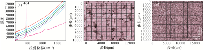
Figure 1.
Raman spectra of (a) quartz at different position under the same experimental conditions and mapping area images and step size ranges of (b) sample 1 and (c) sample 2
-
Figure 2.
Raman spectra of mineral in the samples