
| Professional Committee of Rock and Mineral Testing Technology of the Geological Society of China, National Geological Experiment and Testing Center | Host |
| Citation: |
FANG Biao, YAN Xue-jun, SUN Qing, WU Jing-yi, LI Shu-hua, YAN Jun. Study on the Unique Mineral Microstructure of Seawater Cultured Gray Akoya Pearl by SEM, FTIR and Reflection Spectroscopy[J]. Rock and Mineral Analysis, 2021, 40(1): 42-49. doi: 10.15898/j.cnki.11-2131/td.201908200124

|
Study on the Unique Mineral Microstructure of Seawater Cultured Gray Akoya Pearl by SEM, FTIR and Reflection Spectroscopy
-
Abstract
BACKGROUND Seawater cultured gray Akoya pearls have become popular as jewelry in the recent years. In the early stage, some research focused mainly on investigating the cultured environment of seawater or freshwater pearls, element occurrence characteristics of each structural unit, irradiation treatment and the identification method of irradiated pearls.
OBJECTIVES To further study the gemological characteristics and fine microstructure of a type of gray pearl with a white nucleus.
METHODS Ultraviolet-visible reflection spectrum, micro-infrared spectrum and scanning electron microscope methods were used.
RESULTS A brown transition layer of organic matter between the nacre and nucleus was discovered, which measures several microns in thickness. A layer with no fixed morphology composed of calcite and vaterite in the nacre near the brown transition layer was also discovered. Quasi plates of aragonite exist in the nacre near the surface of the pearl. The morphology of these aragonite tablets in the middle area of the nacre was more regular, the thickness of individual aragonite plate gradually decreased in the direction from the nucleus to the surface of the pearls. The reflectance spectrum of the entire pearl surface was consistent with the spectral characteristics of the outer single nacre. The brown transition layer had no direct effect on the UV-Vis reflectance spectrum of the entire pearl. Therefore, whether or not the brown transition layer affected the gray appearance of the pearl needs further discussion.
CONCLUSIONS The research work has important guiding significance for the coloring mechanism of gray Akoya pearls and the identification of the formation attributes. It can also aid in the recognition of the fine structure and mineralization characteristics of pearls with a thin layer of nacre of 0.3mm to 0.6mm.
-

-
References
[1] 张蓓莉. 系统宝石学[M]. 北京: 地质出版社, 1997. Zhang B L. Systematic gemmology[M]. Beijing: Geological Publishing House, 1997. [2] Kripa V, Mohamed K S, Appukuttan K K, et al. Production of Akoya pearls from the southwest coast of India[J]. Aquaculture, 2007, 262(2): 347-354. [3] Otter L M, Agbaje O B A, Huong L T, et al. Akoya cul-tured pearl farming in eastern Australia[J]. Gems & Gemology, 2017, 53(4): 423-437. [4] Tsujii T. The change of pearl colors by the irradiation with γ-ray or neutron ray[J]. Journal of Radiation Research, 1963, 4(2-4): 120-125. doi: 10.1269/jrr.4.120 [5] 李立平, 陈钟惠. 养殖珍珠的辐照处理[J]. 宝石与宝石学, 2002, 4(3): 16-21. Li L P, Chen Z H. Irradiation treatment of cultured pearls[J]. Journal of Gems and Gemmology, 2002, 4(3): 16-21. [6] Kim H Y, Hanifehpour Y, Narayan A, et al. Structural studies and optical properties of pearl nucleus irradiated by γ-ray[J]. Radiation Effects and Defects in Solids, 2013, 168(9): 696-704. doi: 10.1080/10420150.2012.761997 [7] Kim Y, Choi H, Lee B, et al.Identification of irradiated south sea cultured pearls using electron spin resonance spectroscopy[J].Gems & Gemology, 48(4): 292-299. [8] Choi H, Lee B, Kim Y. Detection of gamma irradiated South Sea cultured pearls[J]. Journal of the Korean Crystal Growth and Crystal Technology, 2012, 22(1): 36-41. doi: 10.6111/JKCGCT.2012.22.1.036 [9] 宋彦军, 张义丞, 武云龙, 等. 银灰色马氏贝海水珍珠的光谱学特征与颜色成因[J]. 矿物学报, 2017, 37(6): 712-716. Song Y J, Zhang Y C, Wu Y L, et al. Spectra characteristics and coloration mechanism of silver-gray color seawater cultured pearls produced by Pinctada Martensii[J]. Acta Mineralogica Sinica, 2017, 37(6): 712-716. [10] 邵惠萍, 严雪俊, 严俊, 等. 应用傅里叶变换红外光谱与紫外可见吸收光谱鉴别两类海水养殖灰色珍珠[J]. 岩矿测试, 2019, 38(5): 489-496. Shao H P, Yan X J, Yan J, et al. Identification of two kinds of seawater cultured gray pearls by Fourier transform infrared spectroscopy and ultraviolet-visible absorption spectroscopy[J]. Rock and Mineral Analysis, 2019, 38(5): 489-496. [11] Ma H Y, Su A A, Zhang B L, et al. Vaterite or aragonite observed in the prismatic layer of freshwater-cultured pearls from South China[J]. Progress in Natural Science, 2009, 19: 817-820. doi: 10.1016/j.pnsc.2008.11.005 [12] Alberto P H, Cuif J P, Dauphin Y, et al. Crystallography of calcite in pearls[J]. European Journal of Mineralogy, 2014, 26(4): 507-516. doi: 10.1127/0935-1221/2014/0026-2390 [13] Ma H Y, Li R K, Yang L X, et al. A modified integrated model of the internal structure of Chinese cultured pearls[J]. Journal of Wuhan University of Technology (Material Science), 2011, 26(3): 510-514. doi: 10.1007/s11595-011-0258-5 [14] Murr L E, Ramirez D A. The microstructure of the cul-tured freshwater pearl[J]. Journal of the Minerals, Metals & Materials Society, 2012, 64(4): 469-474. doi: 10.1007/s11837-012-0297-1 [15] Satitkune S, Monarumit N, Boonmee C, et al. Combina-tion of FTIR and SEM for identifying freshwater-cultured pearls from different quality[J]. Optikai Spektroskopiya, 2016, 120(3): 500-504. doi: 10.1134/S0030400X16030231 [16] Zuo S C, Wei Y G. Microsturcture observation and mechanical behavior modeling for limnetic nacre[J]. Acta Mechanica Sinica, 2008, 24(1): 83-89. doi: 10.1007/s10409-007-0125-y [17] 闻辂. 矿物红外光谱[M]. 重庆: 重庆大学出版社, 1988. Wen L. Mineral infrared spectroscopy[M]. Chongqing: Chongqing University Press, 1988. [18] 张刚生, 李浩璇. 生物成因文石与无机成因文石的FTIR光谱区别[J]. 矿物岩石, 2006, 26(1): 1-4. Zhang G S, Li H X. The FTIR spectra difference between biogenic and abiogenic aragonites[J]. Journal of Mineralogy and Petrology, 2006, 26(1): 1-4. [19] Pokroy B, Fieramosca J S, von Dreele R B, et al. Atomic structure of biogenic aragonite[J]. Chemistry Materials, 2007, 19(13): 3244-3251. doi: 10.1021/cm070187u [20] 张刚生, 丁世磊, 贾太轩, 等. 珍珠及贝壳珍珠层文石的异常红外光谱特征[J]. 宝石和宝石学杂志, 2005, 7(3): 7-9. Zhang G S, Ding S L, Jia T S, et al. Unusual characteristics of FTIR spectra aragonites from nacreous layers of pearls and bivalve shells[J]. Journal of Gems and Gemmology, 2005, 7(3): 7-9. [21] 丁世磊, 张刚生. 天然文石质陶瓷三角帆蚌贝壳的FTIR光谱研究[J]. 光谱学与光谱分析, 2006, 26(12): 2200-2202. Ding S L, Zhang G S. FTIR spectroscopic study on natural aragonite ceramics bivalve shells of Hyriopsis cumingii[J]. Spectroscopy and Spectral Analysis, 2006, 26(12): 2200-2202. [22] Elen S. Update on the identification of treated "Golden" South Sea cultured pearls[J]. Gems & Gemology, 2002, 38(2): 156-159. [23] 史凌云, 郭守国, 王以群. 黑色海水珍珠与人工处理黑色珍珠的光谱学特征研究[J]. 激光与光电子学报, 2012, 49(6): 063002-1-063002-4. Shi L Y, Guo S G, Wang Y Q. Study on spectral characteristics of black saltwater pearls and treated black pearls[J]. Laser & Optoelectronics Progress, 2012, 49(6): 063002-1-063002-4. [24] 亓利剑, 黄艺兰, 曾春光, 等. 各类金色海水珍珠的呈色属性及UV-Vis的反射光谱[J]. 宝石与宝石学, 2008, 10(4): 1-8. Qi L J, Huang Y L, Zeng C G. Colouration attributes and UV-Vis reflection spectra of various golden seawater cultured pearls[J]. Journal of Gems and Gemmology, 2008, 10(4): 1-8. [25] 郭倩, 徐志. 天然金珍珠和染色金珍珠的致色因素和鉴定分析方法研究进展[J]. 岩矿测试, 2015, 34(5): 512-519. Guo Q, Xu Z. Coloring factors of natural and dyed golden pearls and research progress on their identification methods[J]. Rock and Mineral Analysis, 2015, 34(5): 512-519. [26] 陈育, 郭守国, 史凌云. 光谱学在金黄色海水珍珠鉴定中的应用[J]. 光学学报, 2009, 29(6): 1706-1709. Chen Y, Guo S G, Shi L Y. Application of spectroscopy in identification of golden saltwater pearl[J]. Acta Optica Sinica, 2009, 29(6): 1706-1709. [27] Wang W Y, Scarratt K, Hyatt A, et al. Identification of "Chocolate Pearls" treated by ballerina pearl Co[J]. Gems & Gemology, 2006, 42(4): 222-235. [28] Yan J, Zhang J, Tao J B, et al. Origin of the common UV absorption feature in cultured pearls and shells[J]. Journal of Materials Science, 2017, 52(14): 8362-8369. doi: 10.1007/s10853-017-1111-9 [29] Agatonovic K S, Morton D W. The use of UV-visible re-flectance spectroscopy as an objective tool to evaluate pearl quality[J]. Marine Drugs, 2012, 10(7): 1459-1475. [30] 严雪俊, 严俊, 方飚, 等. 钻石的紫外-可见-近红外光谱与光致发光光谱温敏特征及其鉴定指示意义[J]. 光学学报, 2019, 39(9): 0930005-1-0930005-8. Yan X J, Yan J, Fang B, et al. Temperature sensitivity of UV-visible-near infrared and photoluminescence spectra of diamond and its significance for identification[J]. Acta Optica Sinica, 2019, 39(9): 0930005-1-0930005-8. [31] Wang W Y, Ulrika F S, Johansson D H, et al. CVD syn-thetic diamonds from gemesis corp[J]. Gems & Gemology, 2012, 48(2): 80-97. [32] Shigley J E, Breeding C M. Optical defects in diamond: A quick reference chart[J]. Gems & Gemology, 2013, 49(2): 107-111. -
Access History
Figures(4)

Export File
Citation
FANG Biao, YAN Xue-jun, SUN Qing, WU Jing-yi, LI Shu-hua, YAN Jun. Study on the Unique Mineral Microstructure of Seawater Cultured Gray Akoya Pearl by SEM, FTIR and Reflection Spectroscopy[J]. Rock and Mineral Analysis, 2021, 40(1): 42-49. doi: 10.15898/j.cnki.11-2131/td.201908200124
Format
Content
 DownLoad:

-
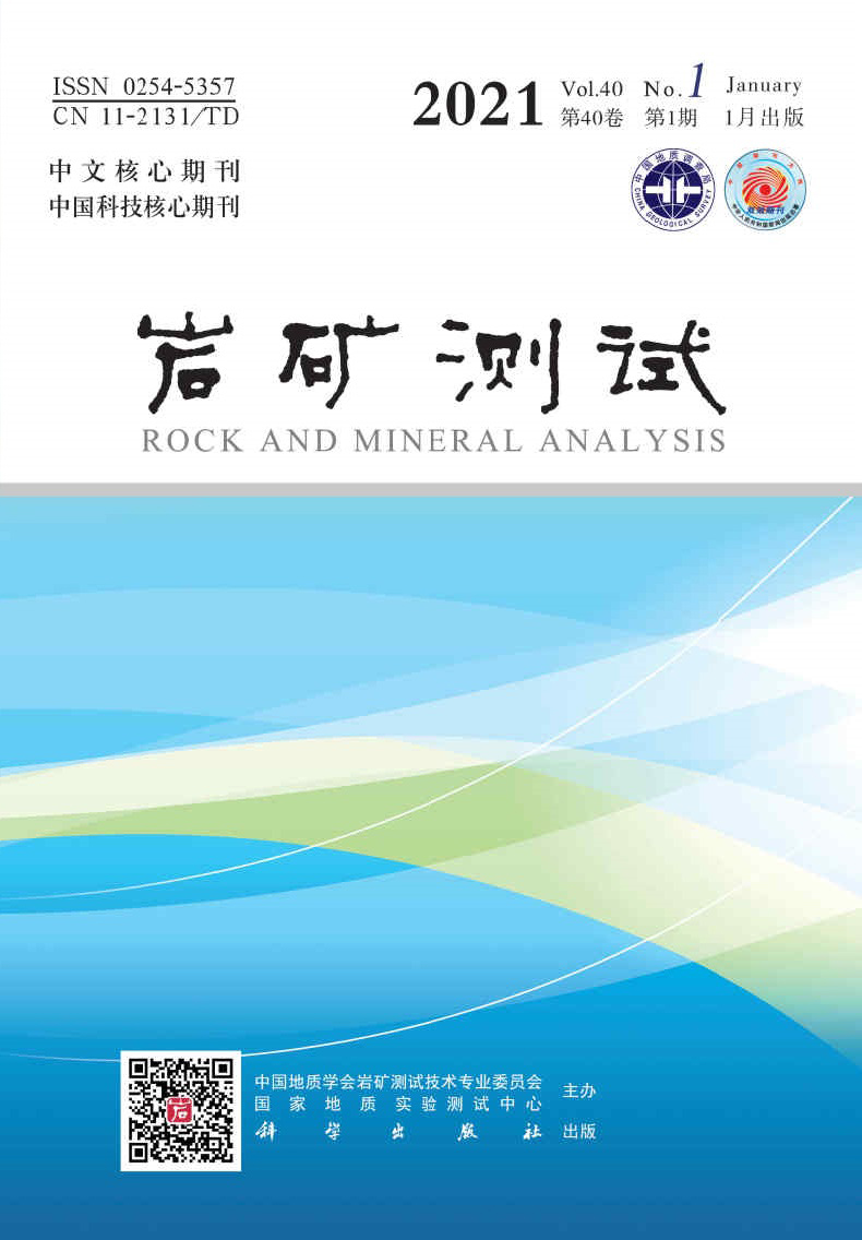
Figure 1.
Optical images of (a) gray pearl with white nucleus and (b) its corresponding structure in the crossing-section
-
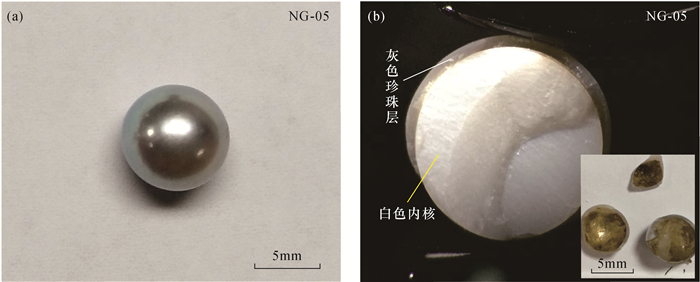
Figure 2.
SEM images of different areas of the pearl from the nucleus to the surface of pearl in the cross-section
-
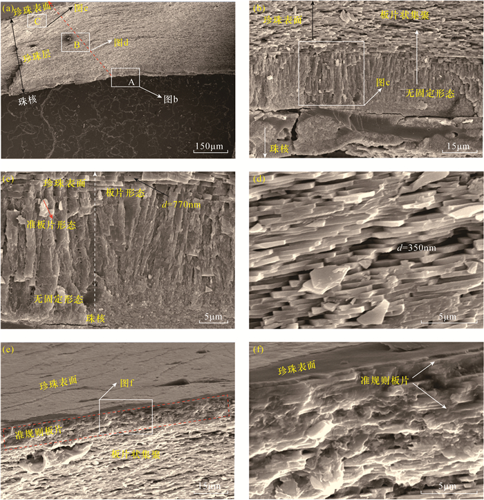
Figure 3.
(a) Optical image, (b) structural schematic diagram of the nacre of gray pearl and (c-f) the corresponding micro-IR spectra of phase composition at different depths under the inner concave surface
-
Figure 4.
UV-Vis reflectance spectra of typical gray pearls with white nucleus and their corresponding nacreous layers covered or uncovered brown organic layer