
| Professional Committee of Rock and Mineral Testing Technology of the Geological Society of China, National Geological Experiment and Testing Center | Host |

| Citation: |
Bing-fei YANG, An-sheng FENG. Study on Accuracy of Quantitative Analysis of Minerals under Rotating Stage Microscopy[J]. Rock and Mineral Analysis, 2018, 37(3): 292-297. doi: 10.15898/j.cnki.11-2131/td.201709270156

|
Study on Accuracy of Quantitative Analysis of Minerals under Rotating Stage Microscopy
-
Abstract
Mineral quantitative analysis by optical microscope is simple and reliable, and has been widely used in process mineralogy and rock-mineral determination. The line measurement method is fast and efficient, and is suitable for fine-grained mineral particles, which plays an important role in the application. However, the conventional test line has the characteristics of directional identity, and cannot deal with the problem of 'preferential orientation' of the analyzed mineral particles. To solve this problem, a new mineral quantitative method by rotating the platform of an optical microscope was conducted. The main principle is that the length ratio of the circular cutting lines of different mineral particles is equal to their volume ratio. The main test process of the method is as follows:rotating the objective table for a circle and testing the angles (ΔK) that the circular cutting line contained, which is intersected by the mineral particles and the visual field under the microscope. By testing a certain number of visual fields (N) and calculating weight percentage (W), the percent content is acquired by the equation W=[∑ΔK/(N×360)]·(δ/Δ)×100%. After theoretical analysis, the galena, sphalerite, and magnetite thin sections were examined. The results show that the new mineral quantitative method is simple, accurate, and the difference value of the test results is less than ±5% compared with the conventional line measurement method. In addition, this method can be used to effectively solve the negative effect of 'preferential orientation' on the test results.-
Keywords:
- rotate the objective table /
- microscope /
- mineral quantitation /
- accuracy
-

-
References
[1] Baum W.Ore characterization, process mineralogy and lab automation a roadmap for future mining[J].Minerals Engineering, 2014, 60:69-73. doi: 10.1016/j.mineng.2013.11.008 [2] Lotter N O, Kormos L J, Oliveira J, et al.Modern process mineralogy:Two case studies[J].Minerals Engineering, 2011, 24:638-650. doi: 10.1016/j.mineng.2011.02.017 [3] 黄凌云, 杨波, 童雄.贵州某铅锌尾矿工艺矿物学研究[J].昆明理工大学学报(自然科学版), 2017, 42(4):25-37. Huang L Y, Yang B, Tong X.Process mineralogy of lead-zinc tailings in Guizhou Province[J].Journal of Kunming University of Science and Technology (Natural Science Edition), 2017, 42(4):25-37. [4] 蒋先强, 熊文良, 曾令熙.国外某铁尾矿中稀土赋存状态研究[J].稀土, 2016, 37(6):32-38. Jiang X Q, Xiong W L, Zeng L X.Study on low grade rare earth occurrences in a foreign iron tailings[J].Chinese Rare Earths, 2016, 37(6):32-38. [5] Zheng X, Yan L, Shuang L, et al.The characteristics study of sphalerite tailings by using MLA[J].Procedia Engineering, 2015, 102:278-286. doi: 10.1016/j.proeng.2015.01.144 [6] 杜谷, 王坤阳, 冉敬, 等.红外光谱/扫描电镜等现代大型仪器岩石矿物鉴定技术及其应用[J].岩矿测试, 2014, 33(9):625-632. Du G, Wang K Y, Ran J, et al.Application of IR/SEM and other modern instruments for mineral identification[J].Rock and Mineral Analysis, 2014, 33(9):625-632. [7] Dirk S. Method Development in Automated Mineralogy[D]. Freiberg: TU Bergakademie, 2015. Method Development in Automated Mineralogy [8] Lotter N O.Modern process mineralogy:An integrated multi-disciplined approach to flowsheeting[J].Minerals Engineering, 2011, 24:1229-1237. doi: 10.1016/j.mineng.2011.03.004 [9] 赵海波, 黄俊玮, 马驰, 等.河南某钨钼矿石工艺矿物学研究[J].金属矿山, 2016(9):122-126. Zhao H B, Huang J W, Ma C, et al.Process mineralogy research of tungsten-molybdenum ore in Henan[J]. Metal Mine, 2016(9):122-126. [10] 马驰, 王守敬, 海东婧, 等.内蒙古赵井沟钽铌矿工艺矿物学研究[J].矿产保护与利用, 2017(6):75-78. Ma C, Wang S J, Hai D J, et al.Process mineralogy of the Zhaojinggou tantalum-niobium ore deposit in Inner Mongolia Province[J].Conservation and Utilization of Mineral Resources, 2017(6):75-78. [11] 周姣花, 汪建宇, 顾茗心, 等.利用X射线衍射和岩矿鉴定等技术研究河南汤家坪钼矿区主要矿物标型特征[J].岩矿测试, 2015, 34(1):82-90. Zhou J H, Wang J Y, Gu M X, et al.The main mineral typomorphic characteristics of the Henan Tangjiaping molybdenum district using X-ray diffraction and rock mineral identification technology[J].Rock and Mineral Analysis, 2015, 34(1):82-90. [12] 彭艳华, 彭光菊, 贾利攀, 等.湖南宝山铅锌矿西部矿带银的工艺矿物学研究[J].岩矿测试, 2013, 32(5):729-737. Peng Y H, Peng G J, Jia L P, et al.Technological mineralogy research of silver in the lead-zinc ore deposit in West Baoshan, Hunan Province[J].Rock and Mineral Analysis, 2013, 32(5):729-737. [13] Goodwin P C, Johnson B, Frevert C W.Microscopy, Im-muno-histochemistry, Digital Imaging, and Quantitative Microscopy[M].Elsevier Press, 2018:53-66. [14] Ueda T, Oki T, Koyanaka S.Stereological correction me-thod based on sectional texture analysis for the liberation distribution of binary particle systems[J].Advanced Powder Technology, 2017, 28:1391-1398. doi: 10.1016/j.apt.2017.03.007 [15] Ueda T, Oki T, Koyanaka S.Stereological bias for sph-erical particles with various particle compositions[J].Advanced Powder Technology, 2016, 27:1828-1838. doi: 10.1016/j.apt.2016.06.016 [16] de Souza D S, da Silva Assis W L, Rios P R, et al.Ste-reological analysis of the microstructure of pure iron with random nucleation[J].Journal of Materials Research and Technology, 2014, 3(4):349-353. doi: 10.1016/j.jmrt.2014.08.002 -
Access History
Figures(4)
Tables(1)

Export File
Citation
Bing-fei YANG, An-sheng FENG. Study on Accuracy of Quantitative Analysis of Minerals under Rotating Stage Microscopy[J]. Rock and Mineral Analysis, 2018, 37(3): 292-297. doi: 10.15898/j.cnki.11-2131/td.201709270156
Format
Content
 DownLoad:

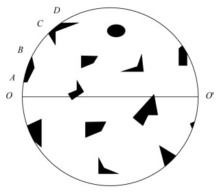
- Figure 1. The sketch of visual field under the microscope
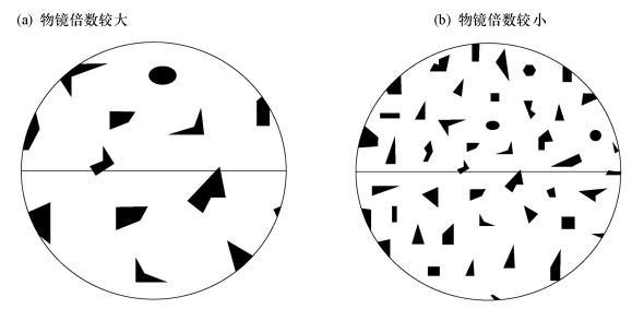
- Figure 2. The sketchs of visual field under different magnification of objective
- Figure 3. Moving way of the visual field under the microscope
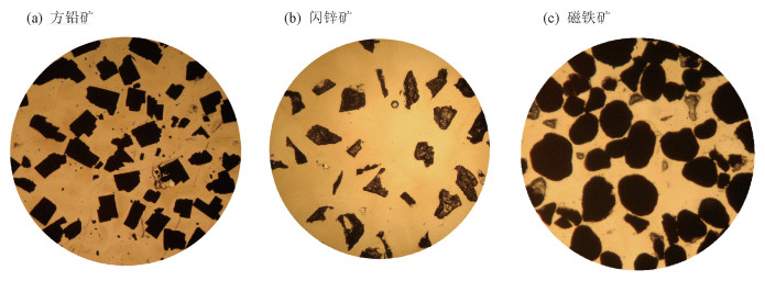
- Figure 4. Visual fields under the microscope