
| Professional Committee of Rock and Mineral Testing Technology of the Geological Society of China, National Geological Experiment and Testing Center | Host |
| Citation: |
TAO Peng, XIE Shiwen, LONG Tao, MA Mingzhu, CHE Xiaochao. Atom Probe Tomography (APT) and Its Application in Ore Deposits[J]. Rock and Mineral Analysis, 2023, 42(5): 957-969. doi: 10.15898/j.ykcs.202307300100

|
Atom Probe Tomography (APT) and Its Application in Ore Deposits
-
Abstract
Atom Probe Tomography (APT) is a test analysis technique that provides quantitative three-dimensional element and isotope analysis at subnanometer resolution, with extremely high spatial resolution and low detection limits[13]. Compared with traditional geological analysis techniques, APT has unique technical advantages, which can be used to analyze the elemental composition of minerals <0.0007μm3 in volume[14], reveal the complexity of mineral composition at the nanoscale, and provide a new understanding of the geological evolution process. APT has been in development for over 50 years, and continuous technological advancements have led to its wider application range. At the beginning of APT design, it was only used for conductive materials. From the end of the 20th century to the beginning of the 21st century, the application of laser pulse mode enabled APT to be applied to semiconductors and insulating materials[15-19], and the application of Local Electrode TM Atom Probe (LEAP) improved several key parameters such as the data acquisition rate and mass resolution of APT by several orders of magnitude[20]. At present, most of the geological application work of APT is carried out by LEAP in laser-assisted mode[13]. In recent years, the unique technical advantages of APT have attracted increasing attention in geological research, and their advantages in ore deposit research have become more prominent. Some important research results have been published[21-31]. However, on the whole, its application in ore deposits and even geology is still in its infancy. The development history, basic principle, selection method of area of interest and needle tip sample preparation of APT are briefly introduced in this paper. Based on this, representative application achievements of APT in ore deposit research by domestic and foreign scholars in recent years are collected and summarized. In ore deposit research, APT is mainly applied in three aspects: the occurrence states of ore-forming elements, nanoscale inclusions, and stable isotope composition[21-31]. At present, most research results focus on the analysis of the occurrence status of ore-forming elements, especially pyrite or other minerals with simple chemical composition related to gold deposits. APT has successfully revealed three main occurrence states of ore-forming elements on the atomic scale: uniform distribution, nanoparticle and enrichment at low angle grain boundaries and dislocations[21-25]. For example, gold can be uniformly distributed in the form of dispersed lattice bound gold in the arsenic-rich overgrowth rim of pyrite[21], and can form nanoclusters of different sizes in arsenopyrite[22]. It can also host in the low angle boundary of pyrite related to deformation[24]. In terms of nano inclusions and stable isotope composition, the research mainly focuses on pyrite nano fluid inclusions and S isotopes[26-31]. For example, nano telluride inclusions along pyrite fractures in low-sulfidation type epithermal Au-Ag-Te deposit[26] and the method for obtaining quantitative δ34S measurement value from APT datasets of pyrite[29]. The relevant results are shown in Fig.E.1. So far, the applications of APT in ore deposits research have mainly focused on the occurrence state of ore-forming elements, achieving three-dimensional visualization of atomic scale element distribution that was previously unimaginable, providing a new perspective for people to understand and explain the ore-forming process. In terms of nano inclusions and stable isotope composition, although the applications of APT are not as rich as the former, some important new understandings have been obtained, showing a good application prospect. While APT is rapidly developing in the field of ore deposits, there are still many problems to be solved in its practical application. For example, the extremely small sample volume, time-consuming selection of specific areas, the background noise carried by the mass spectrometry itself, the correct interpretation of complex spectral peaks, and the accuracy of data three-dimensional reconstruction. However, it is foreseeable that with the continuous progress of technology, APT will become more popular and easier to use, increasing numbers of deposit researchers will pay attention to APT, and more ore deposit samples with complex types, structures and chemical compositions will apply this technology for in-depth research, which may change or even completely subvert our understanding of some basic scientific problems in ore deposits.
-

-
References
[1] 李文昌, 李建威, 谢桂青, 等. 中国关键矿产现状, 研究内容与资源战略分析[J]. 地学前缘, 2022, 29(1): 1−13. Li W C, Li J W, Xie G Q, et al. Critical minerals in China: Current status, research focus and resource strategic analysis[J]. Earth Science Frontiers, 2022, 29(1): 1−13. [2] 蒋少涌, 赵葵东, 姜海, 等. 中国钨锡矿床时空分布规律, 地质特征与成矿机制研究进展[J]. 科学通报, 2020, 65(33): 3730−3745. Jiang S Y, Zhao K D, Jiang H, et al. Spatiotemporal distribution, geological characteristics and metallogenic mechanism of tungsten and tin deposits in China: An overview[J]. Chinese Science Bulletin, 2020, 65(33): 3730−3745. [3] 涂家润, 卢宜冠, 孙凯, 等. 应用微束分析技术研究铜钴矿床中钴的赋存状态[J]. 岩矿测试, 2022, 41(2): 226−238. doi: 10.15898/j.cnki.11-2131/td.202112060194 Tu J R, Lu Y G, Sun K, et al. Application of microbeam analytical technology to study the occurrence of cobalt from copper-cobalt deposits[J]. Rock and Mineral Analysis, 2022, 41(2): 226−238. doi: 10.15898/j.cnki.11-2131/td.202112060194 [4] 侯增谦, 陈骏, 翟明国. 战略性关键矿产研究现状与科学前沿[J]. 科学通报, 2020, 65(33): 3651−3652. doi: 10.1360/TB-2020-1417 Hou Z Q, Chen J, Zhai M G. Current status and frontiers of research on critical mineral resources[J]. Chinese Science Bulletin, 2020, 65(33): 3651−3652. doi: 10.1360/TB-2020-1417 [5] 翟明国, 吴福元, 胡瑞忠, 等. 战略性关键金属矿产资源: 现状与问题[J]. 中国科学基金, 2019, 33(2): 106−111. Zhai M G, Wu F Y, Hu R Z, et al. Critical metal mineral resources: Current research status and scientific issues[J]. Bulletin of National Natural Science Foundation of China, 2019, 33(2): 106−111. [6] London D. Rare-element granitic pegmatites[J]. Reviews in Economic Geology, 2016, 18: 165−194. [7] 涂光炽, 高振敏, 胡瑞忠, 等. 分散元素地球化学及成矿机制[M]. 北京: 地质出版社, 2004: 1-424. Tu G Z, Gao Z M, Hu R Z, et al. The Geochemistry and Ore-forming Mechanism of the Dispersed Elements[M]. Beijing: Geological Publishing House, 2004: 1-424. [8] 李超, 王登红, 屈文俊, 等. 关键金属元素分析测试技术方法应用进展[J]. 岩矿测试, 2020, 39(5): 658−669. doi: 10.15898/j.cnki.11-2131/td.201907310115 Li C, Wang D H, Qu W J, et al. A review and perspective on analytical methods of critical metal elements[J]. Rock and Mineral Analysis, 2020, 39(5): 658−669. doi: 10.15898/j.cnki.11-2131/td.201907310115 [9] Chen L L, Chen Y, Feng X X, et al. Uranium occurrence state in the Tarangaole area of the Ordos Basin, China: Implications for enrichment and mineralization[J]. Ore Geology Reviews, 2019, 115: 103034. doi: 10.1016/j.oregeorev.2019.103034 [10] 员媛娇, 范成龙, 吕喜平, 等. 电子探针和 LA-ICP-MS 技术研究内蒙古浩尧尔忽洞金矿床毒砂矿物学特征[J]. 岩矿测试, 2022, 41(2): 211−225. Yun Y J, Fan C L, Lyu X P, et al. Application of EPMA and LA-ICP-MS to study mineralogy of arsenopyrite from the Haoyaoerhudong gold deposit, Inner Mongolia, China[J]. Rock and Mineral Analysis, 2022, 41(2): 211−225. [11] Deol S, Deb M, Large R R, et al. LA-ICPMS and EPMA studies of pyrite, arsenopyrite and loellingite from the Bhukia—Jagpura gold prospect, Southern Rajasthan, India: Implications for ore genesis and gold remobilization[J]. Chemical Geology, 2012, 326: 72−87. [12] 汪超, 王瑞廷, 刘云华, 等. 陕西商南三官庙金矿床地质特征, 金的赋存状态及矿床成因探讨[J]. 矿床地质, 2021, 40(3): 491−508. Wang C, Wang R T, Liu Y H, et al. Geological characteristics, modes of occurrence of gold and genesis of San’guanmiao gold deposit, Shangnan, Shaanxi Province[J]. Mineral Deposits, 2021, 40(3): 491−508. [13] Reddy S M, Saxey D W, Rickard W D A, et al. Atom probe tomography: Development and application to the geosciences[J]. Geostandards and Geoanalytical Research, 2020, 44(1): 5−50. doi: 10.1111/ggr.12313 [14] Fougerouse D, Kirkland C L, Saxey D W, et al. Nanoscale isotopic dating of monazite[J]. Geostandards and Geoanalytical Research, 2020, 44(4): 637−652. doi: 10.1111/ggr.12340 [15] Perea D E, Lensch J L, May S J, et al. Composition analysis of single semiconductor nanowires using pulsed-laser atom probe tomography[J]. Applied Physics A, 2006, 85: 271−275. doi: 10.1007/s00339-006-3710-1 [16] Larson D J, Alvis R L, Lawrence D F, et al. Analysis of bulk dielectrics with atom probe tomography[J]. Microscopy and Microanalysis, 2008, 14(S2): 1254−1255. doi: 10.1017/S1431927608083657 [17] Bachhav M, Danoix R, Danoix F, et al. Investigation of wüstite (Fe1- x O) by femtosecond laser assisted atom probe tomography[J]. Ultramicroscopy, 2011, 111(6): 584−588. doi: 10.1016/j.ultramic.2010.11.023 [18] Pérez-Huerta A, Laiginhas F, Reinhard D A, et al. Atom probe tomography (APT) of carbonate minerals[J]. Micron, 2016, 80: 83−89. doi: 10.1016/j.micron.2015.10.001 [19] Larson D J, Prosa T J, Perea D E, et al. Atom probe tomography of nanoscale electronic materials[J]. Mrs Bulletin, 2016, 41: 30−34. doi: 10.1557/mrs.2015.308 [20] Ulfig R M, Larson D J, Kelly T F, et al. Performance advances in LEAP systems[J]. Microscopy and Microanalysis, 2014, 20(S3): 1120−1121. doi: 10.1017/S1431927614007338 [21] Gopon P, Douglas J O, Auger M A, et al. A nanoscale investigation of Carlin-type gold deposits: An atom-scale elemental and isotopic perspective[J]. Economic Geology, 2019, 114(6): 1123−1133. doi: 10.5382/econgeo.4676 [22] Fougerouse D, Reddy S M, Saxey D W, et al. Nanoscale gold clusters in arsenopyrite controlled by growth rate not concentration: Evidence from atom probe microscopy[J]. American Mineralogist, 2016, 101(8): 1916−1919. doi: 10.2138/am-2016-5781CCBYNCND [23] Fougerouse D, Cugerone A, Reddy S M, et al. Nanoscale distribution of Ge in Cu-rich sphalerite[J]. Geochimica et Cosmochimica Acta, 2023, 346: 223−230. doi: 10.1016/j.gca.2023.02.011 [24] Fougerouse D, Reddy S M, Aylmore M, et al. A new kind of invisible gold in pyrite hosted in deformation-related dislocations[J]. Geology, 2021, 49(10): 1225−1229. doi: 10.1130/G49028.1 [25] Dubosq R, Rogowitz A, Schweinar K, et al. A 2D and 3D nanostructural study of naturally deformed pyrite: Assessing the links between trace element mobility and defect structures[J]. Contributions to Mineralogy and Petrology, 2019, 174(9): 72. doi: 10.1007/s00410-019-1611-5 [26] Börner F, Keith M, Fougerouse D, et al. Between defects and inclusions: The fate of tellurium in pyrite[J]. Chemical Geology, 2023: 121633. [27] Dubosq R, Rogowitz A, Schneider D A, et al. Fluid inclusion induced hardening: Nanoscale evidence from naturally deformed pyrite[J]. Contributions to Mineralogy and Petrology, 2021, 176(2): 15. doi: 10.1007/s00410-021-01774-9 [28] Dubosq R, Gault B, Hatzoglou C, et al. Analysis of nanoscale fluid inclusions in geomaterials by atom probe tomography: Experiments and numerical simulations[J]. Ultramicroscopy, 2020, 218: 113092. doi: 10.1016/j.ultramic.2020.113092 [29] Gopon P, Douglas J O, Meisenkothen F, et al. Atom probe tomography for isotopic analysis: Development of the 34S/32S system in sulfides[J]. Microscopy and Microanalysis, 2022, 28(4): 1127−1140. doi: 10.1017/S1431927621013568 [30] Lewis J B, Isheim D, Floss C, et al. 12C/13C-ratio determination in nanodiamonds by atom-probe tomography[J]. Ultramicroscopy, 2015, 159: 248−254. doi: 10.1016/j.ultramic.2015.05.021 [31] Darling J R, White L F, Kizovski T, et al. The shocking state of apatite and merrillite in shergottite Northwest Africa 5298 and extreme nanoscale chlorine isotope variability revealed by atom probe tomography[J]. Geochimica et Cosmochimica Acta, 2021, 293: 422−437. doi: 10.1016/j.gca.2020.11.007 [32] 王碧雯, 李秋立. 原子探针工作原理及其在地球科学中的应用[J]. 矿物岩石地球化学通报, 2020, 39(6): 1108−1118. doi: 10.19658/j.issn.1007-2802.2020.39.101 Wang B W, Li Q L. An introduction to principle of atom probe and its applications in Earth sciences[J]. Bulletin of Mineralogy, Petrology and Geochemistry, 2020, 39(6): 1108−1118. doi: 10.19658/j.issn.1007-2802.2020.39.101 [33] Gault B, Chiaramonti A, Cojocaru-Mirédin O, et al. Atom probe tomography[J]. Nature Reviews Methods Primers, 2021, 1(1): 51. doi: 10.1038/s43586-021-00047-w [34] Saxey D W, Moser D E, Piazolo S, et al. Atomic worlds: Current state and future of atom probe tomography in geoscience[J]. Scripta Materialia, 2018, 148: 115−121. doi: 10.1016/j.scriptamat.2017.11.014 [35] Wu Y F, Fougerouse D, Evans K, et al. Gold, arsenic, and copper zoning in pyrite: A record of fluid chemistry and growth kinetics[J]. Geology, 2019, 47(7): 641−644. doi: 10.1130/G46114.1 [36] Miller M K, Russell K F, Thompson K, et al. Review of atom probe FIB-based specimen preparation methods[J]. Microscopy and Microanalysis, 2007, 13(6): 428−436. doi: 10.1017/S1431927607070845 [37] Hough R M, Noble R R P, Reich M. Natural gold nanoparticles[J]. Ore Geology Reviews, 2011, 42(1): 55−61. doi: 10.1016/j.oregeorev.2011.07.003 [38] McLeish D F, Williams-Jones A E, Vasyukova O V, et al. Colloidal transport and flocculation are the cause of the hyperenrichment of gold in nature[J]. Proceedings of the National Academy of Sciences, 2021, 118(20): e2100689118. doi: 10.1073/pnas.2100689118 [39] Petrella L, Thébaud N, Fougerouse D, et al. Nanoparticle suspensions from carbon-rich fluid make high-grade gold deposits[J]. Nature Communications, 2022, 13(1): 3795. doi: 10.1038/s41467-022-31447-5 [40] Cabri L J, Chryssoulis S L, de Villiers J P R, et al. The nature of “invisible” gold in arsenopyrite[J]. The Canadian Mineralogist, 1989, 27(3): 353−362. [41] Palenik C S, Utsunomiya S, Reich M, et al. “Invisible” gold revealed: Direct imaging of gold nanoparticles in a Carlin-type deposit[J]. American Mineralogist, 2004, 89(10): 1359−1366. doi: 10.2138/am-2004-1002 [42] Cook N J, Ciobanu C L, Pring A, et al. Trace and minor elements in sphalerite: A LA-ICPMS study[J]. Geochimica et Cosmochimica Acta, 2009, 73(16): 4761−4791. doi: 10.1016/j.gca.2009.05.045 [43] Johan Z. Indium and germanium in the structure of sphalerite: An example of coupled substitution with copper[J]. Mineralogy and Petrology, 1988, 39: 211−229. doi: 10.1007/BF01163036 [44] 韩英, 王京彬, 祝新友, 等. 广东凡口铅锌矿床流体包裹体特征及地质意义[J]. 矿物岩石地球化学通报, 2013(1): 81−86. Han Y, Wang J B, Zhu X Y, et al. The characteristics and its geological significance of the fluid inclusion in the Fankou lead-zinc deposit, Guangdong[J]. Bulletin of Mineralogy, Petrology and Geochemistry, 2013(1): 81−86. [45] 倪培, 范宏瑞, 丁俊英. 流体包裹体研究进展[J]. 矿物岩石地球化学通报, 2014, 33(1): 1−5. doi: 10.19658/j.issn.1007-2802.2021.40.056 Ni P, Fan H R, Ding J Y. Progress in fluid inclusions[J]. Bulletin of Mineralogy, Petrology and Geochemistry, 2014, 33(1): 1−5. doi: 10.19658/j.issn.1007-2802.2021.40.056 [46] 李晓东, 张艳, 韩润生, 等. 流体包裹体研究进展及其在矿床学中的应用[J]. 地质论评, 2022, 68(6): 14. doi: 10.16509/j.georeview.2022.07.065 Li X D, Zhang Y, Han R S, et al. Research progress of fluid inclusions and its application in ore deposit[J]. Geological Review, 2022, 68(6): 14. doi: 10.16509/j.georeview.2022.07.065 [47] Taylor S D, Gregory D D, Perea D E, et al. Pushing the limits: Resolving paleoseawater signatures in nanoscale fluid inclusions by atom probe tomography[J]. Earth and Planetary Science Letters, 2022, 599: 117859. doi: 10.1016/j.jpgl.2022.117859 -
Access History

Figures(9)

Export File
Citation
TAO Peng, XIE Shiwen, LONG Tao, MA Mingzhu, CHE Xiaochao. Atom Probe Tomography (APT) and Its Application in Ore Deposits[J]. Rock and Mineral Analysis, 2023, 42(5): 957-969. doi: 10.15898/j.ykcs.202307300100
Format
Content
 DownLoad:


-
Figure 1.
Comparison of analysis volumes for common microanalysis techniques. Left side panel: each volume is represented on a schematic monazite grain (diagram to scale). APT is capable of measuring the isotopic compositions of minerals at the nanoscale for volumes <0.0007μm3 (Modified from Fougerouse, et al[14]).
-
Figure 2.
Schematic diagram of working principle and analysis results of APT.
-
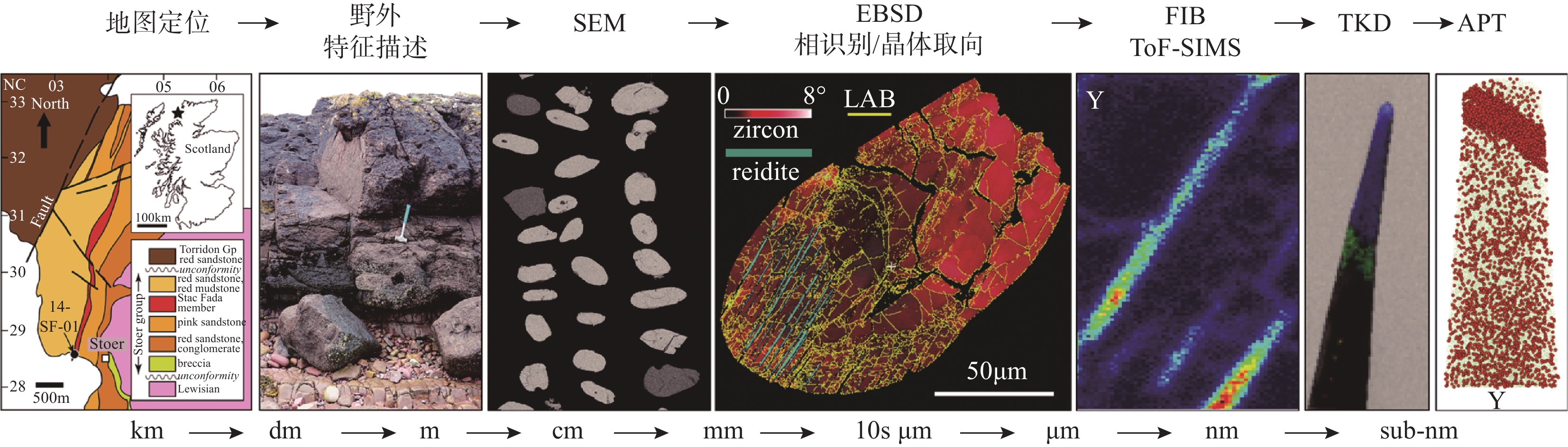
Figure 3.
Characterization workflow of geological samples before APT analysis. Illustrated workflow starts with geological mapping and progresses through high spatial resolution techniques (Modified from Reddy, et al[13]).
-
Figure 4.
Processes for the preparation of APT tip sample (Modified from Gault, et al[33]).
-
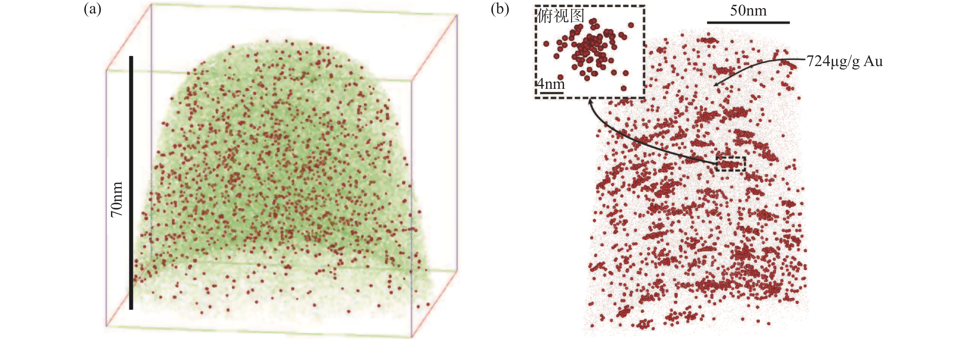
Figure 5.
Structurally bound Au and discrete nanoparticles of Au.
-
Figure 6.
Different occurrence states of Ge in sphalerite (Modified from Fougerouse, et al[23]).
-
Figure 7.
Nanoscale imaging of low-angle boundary (Modified from Fougerouse, et al)[24].
-
Figure 8.
Nanoscale imaging of fluid inclusions (Modified from Dubosq, et al[27]).
-
Figure E.1.
The working principle of APT and its representative applications in ore deposits research. a—Working principle of APT. Modified from Gault, et al[33]; b—APT 3D atom map of the Au in arsenopyrite. Each sphere represents an Au atom. Au atoms are segregated in clusters. The top left illustration is 5nm slice through the largest Au cluster. Modified from Fougerouse, et al[22]; c—Nanoscale imaging of fluid inclusions. Reconstruction of APT specimen displays the distribution of Fe (pink dots), As (turquoise isosurfaces) and O (red isosurfaces) compositions, revealing globular high-density features (fuid inclusions) and one linear feature linking two larger high-density features. Modified from Dubosq, et al[27]; d—APT datasets from R47_01719 chlorapatite. Selected isoconcentration surfaces (ICS) are shown for each whole dataset (Fe=blue, Cl=green, Mn=red). Modified from Darling, et al[31].