
| Professional Committee of Rock and Mineral Testing Technology of the Geological Society of China, National Geological Experiment and Testing Center | Host |
| Citation: |
WANG Kunyang, DU Gu, WANG Guan, HE Jiale. The Morphological Characteristics of Clay Minerals in a Tight Sandstone Reservoir by Atomic Force Microscopy[J]. Rock and Mineral Analysis, 2025, 44(2): 245-253. doi: 10.15898/j.ykcs.202404040075

|
The Morphological Characteristics of Clay Minerals in a Tight Sandstone Reservoir by Atomic Force Microscopy
-
Abstract
Clay minerals, as one of the main components of unconventional oil and gas reservoirs, have significant implications for the evaluation of unconventional oil and gas reservoirs in terms of their fine characterization of nano/sub-nano morphological features. By using atomic force microscopy (AFM) micro-area analysis technology, the problem of secondary modification of nano-pore structure caused by the conductive film in the pretreatment process of electron microscopy was solved; it made up for the defect that electron microscopy requires the sample to be conductive and can directly observe the morphological characteristics of the sample. Here, it was observed through AFM, that in the late diagenetic stage of the tight sandstone of the Xujiahe Formation in western Sichuan, some clay minerals developed parallel stepped stripes, with a large number of nano-pores formed at the concave angles on both sides of the steps, which were the main components of inorganic pores. Secondly, clay minerals had the same diagenetic evolution sequence, but their crystal morphologies were different, indicating that there was a spatial coupling relationship between their morphological features and diagenesis. The BRIEF REPORT is available for this paper at
http://www.ykcs.ac.cn/en/article/doi/10.15898/j.ykcs.202404040075 . -

-
References
[1] 叶荣, 涂光炽, 马喆生, 等. 热液矿床矿物微形貌与晶体生长环境研究[J]. 地学前缘, 2005, 12(2): 240−246. doi: 10.3321/j.issn:1005-2321.2005.02.026 Ye R, Tu G Z, Ma Z S, et al. The surface micromorphology of minerals in hydrothermal ore deposits and growth environments of crystal[J]. Earth Science Frontiers, 2005, 12(2): 240−246. doi: 10.3321/j.issn:1005-2321.2005.02.026 [2] 陈光远, 孙岱生. 成因矿物学找矿矿物学新进展[M]. 北京: 原子能出版社, 1998: 12−16. Chen G Y, Sun D S. New development in genetic mineralogy and prospecting mineralogy[M]. Beijing: Atomic Energy Press, 1998: 12−16. [3] 陈光远, 鲁安怀. 矿物成分标型继承性与金矿矿质来源[J]. 地学前缘, 1994, 1(3−4): 204−209. doi: 10.3321/j.issn:1005-2321.1994.04.025 Chen G Y, Lu A H. Chemical inheritance of mineral typomorphism and source of gold[J]. Earth Science Frontiers, 1994, 1(3−4): 204−209. doi: 10.3321/j.issn:1005-2321.1994.04.025 [4] 王坤阳, 杜谷. 利用原子力显微镜与能谱-扫描电镜研究页岩孔隙结构特征[J]. 岩矿测试, 2020, 39(6): 839−846. doi: 10.15898/j.cnki.11-2131/td.202004180053 Wang K Y, Du G. Study on the pore structure characteristics of shale by atomic force microscope and energy spectrum-scanning electron microscope[J]. Rock and Mineral Analysis, 2020, 39(6): 839−846. doi: 10.15898/j.cnki.11-2131/td.202004180053 [5] 王羽, 汪丽华, 王建强, 等. 基于聚焦离子束-扫描电镜方法研究页岩有机孔三维结构[J]. 岩矿测试, 2018, 37(3): 235−243. doi: 10.15898/j.cnki.11-2131/td.201612210188 Wang Y, Wang L H, Wang J Q, et al. Three-dimension characterization of organic matter pore structures of shale using focused ion beam-scanning electron microscope[J]. Rock and Mineral Analysis, 2018, 37(3): 235−243. doi: 10.15898/j.cnki.11-2131/td.201612210188 [6] 许谊, 徐毓娴, 惠梅, 等. 微分相衬干涉显微镜定量测量表面形貌[J]. 光学精密工程, 2001, 9(3): 227−230. doi: 10.3321/j.issn:1004-924X.2001.03.006 Xu Y, Xu Y X, Hui M, et al. Quantitative surface topography determination by differential interference contrast microscopy[J]. Optics and Precision Engineering, 2001, 9(3): 227−230. doi: 10.3321/j.issn:1004-924X.2001.03.006 [7] Barty A, Nugent K A, Paganin D, et al. Quantitative optical phase microscopy[J]. Optics Letters, 1998, 23(11): 817−819. doi: 10.1364/OL.23.000817 [8] Liao L B, Li S Z. Effect of SPM scanning range on the micromorphology parameters[J]. International Journal of Minerals, Metallurgy and Materials, 1995, 5(1): 36−38. [9] 戚明辉, 李君军, 曹茜. 基于扫描电镜和JMicroVision图像分析软件的泥页岩孔隙结构表征研究[J]. 岩矿测试, 2019, 38(3): 260−269. doi: 10.15898/j.cnki.11-2131/td.201901160008 Qi M H, Li J J, Cao Q. The pore structure characterization of shale based on scanning electron microscopy and JMicroVision[J]. Rock and Mineral Analysis, 2019, 38(3): 260−269. doi: 10.15898/j.cnki.11-2131/td.201901160008 [10] 朱炎铭, 王阳, 陈尚斌, 等. 页岩储层孔隙结构多尺度定性-定量综合表征: 以上扬子海相龙马溪组为例[J]. 地学前缘, 2016, 23(1): 154−163. doi: 10.13745/j.esf.2016.01.01 Zhu Y M, Wang Y, Chen S B, et al. Qualitative-quantitative multiscale characterization of pore structures in shale reservoirs: A case study of Longmaxi Formation in the upper Yangze area[J]. Earth Science Frontiers, 2016, 23(1): 154−163. doi: 10.13745/j.esf.2016.01.01 [11] Klaver J, Desbois G, Urai J L, et al. BIB-SEM study of the pore space morphology in early mature Posidonia shale from the Hils area, Germany[J]. International Journal of Coal Geology, 2012, 103: 12−25. doi: 10.1016/j.coal.2012.06.012 [12] 黄振凯, 陈建平, 王义军, 等. 微米CT在烃源岩微观结构表征方面的应用[J]. 石油实验地质, 2016, 38(3): 418−422. doi: 10.11781/sysydz201603418 Huang Z K, Chen J P, Wang Y J, et al. Application of micron CT in the characterization of microstructure in source rocks[J]. Petroleum Geology & Experiment, 2016, 38(3): 418−422. doi: 10.11781/sysydz201603418 [13] Gratz A J, Hillner P E. Poisoning of calcite crystal growth viewed in the atomic force microscope (AFM)[J]. Journal of Crystal Growth, 1993, 129(3): 789−793. doi: 10.1016/0022-0248(93)90515-X [14] 姚素平, 焦堃, 张科, 等. 煤纳米孔隙结构的原子力显微镜研究[J]. 科学通报, 2011, 56(22): 1820−1827. doi: 10.1360/csb2011-56-22-1820 Yao S P, Jiao K, Zhang K, et al. An atomic force microscopy study of coal nanopore structure[J]. Chinese Science Bulletin, 2011, 56(22): 1820−1827. doi: 10.1360/csb2011-56-22-1820 [15] Hochella M F, Jr Eggleston C M, Elings V B, et al. Mineralogy in two dimensions: Scanning tunneling microscopy of semiconducing minerals with implications for geochemical reactivity[J]. American Mineralogist, 1989, 74: 1233−1246. [16] Friedbacher G, Hansma P K. Imaging powders with the atomic force microscope: From biominerals to commercial materials[J]. Science, 1991, 253: 1261−1262. doi: 10.1126/science.253.5025.1261 [17] Jr Eggleston C M, Hochella M F. Scanning tunneling microscope of sulfide surface[J]. Geochimica et Cosmochimica Acta, 1990, 54: 1551−1517. doi: 10.1016/0016-7037(90)90176-L [18] Johnson P D, Eggleston C M, Hochella M F. Imaging molecular-scale structure and microtopography of hematite with the atomic force microscope[J]. American Mineralogist, 1991, 76: 1442−1445. [19] 廖立兵, 马喆生, 施倪承. 扫描隧道显微镜和原子力显微镜在矿物学研究中的应用现状及前景[J]. 现代地质, 1993, 7(4): 5. Liao L B, Ma Z S, Shi N C. Application status and prospect of scanning tunneling microscope and atomic force microscope in mineralogy research[J]. Geoscience, 1993, 7(4): 5. [20] 孙全力, 孙晗森, 贾趵, 等. 川西须家河组致密砂岩储层绿泥石成因及其优质储层关系[J]. 石油与天然气地质, 2012, 33(5): 751−757. doi: 10.11743/ogg20120512 Sun Q L, Sun H S, Jia B, et al. Genesis of chlorite in tight sandstone reservoirs of Xujiahe Formation in western Sichuan and its relationship with high-quality reservoirs[J]. Oil & Gas Geology, 2012, 33(5): 751−757. doi: 10.11743/ogg20120512 [21] 周张健. 蒙脱石伊利石化的控制因素、转化机制及其转化模型的研究综述[J]. 地质科技情报, 1994, 13(4): 41−46. Zhou Z J. Review on control factors, transformation mechanism and transformation model of montmorillonite illization[J]. Geological Science and Technology Bulletin, 1994, 13(4): 41−46. [22] 李胜荣, 许虹, 申俊峰, 等. 结晶学与矿物学[M]. 北京: 地质出版社, 2008: 9−18. Li S R, Xu H, Shen J F, et al. Crystallography and mineralogy[M]. Beijing: Geological Publishing House, 2008: 9−18. [23] 潘兆橹. 结晶学及矿物学[M]. 北京: 地质出版社, 1993: 15−26. Pan Z L. Crystallography and mineralogy [M]. Beijing: Geological Publishing House, 1993: 15−26. [24] 王文魁, 王继扬, 赵珊茸, 等. 晶体形貌学[M]. 北京: 中国地质大学出版社, 2001: 22−27. Wang W K, Wang J Y, Zhao S R, et al. Crystal morphology[M]. Beijing: China University of Geosciences Press, 2001: 22−27. [25] 张天乐, 王宗良. 中国粘土矿物的电子显微研究[M]. 北京: 地质出版社, 1978: 33−41. Zhang T L, Wang Z L. Electron microscopy of clay minerals in China [M]. Beijing: Geological Publishing House, 1978: 33−41. [26] 徐春华, 朱光, 刘国生, 等. 伊利石结晶度在恢复地层剥蚀量中的应用—伊利石结晶度在恢复地层剥蚀量中的应用——以合肥盆地安参1井白垩系剥蚀量的恢复为例[J]. 地质科技情报, 2005, 24(1): 41−44. Xu C H, Zhu G, Liu G S, et al. Application of crystallinity of illite to recover denudation quantity: An example of Cretaceous denudation quantity recovery of Well Ancan 1 in Hefei Basin,Anhui Province[J]. Geological Science and Technology, 2005, 24(1): 41−44. [27] 张有瑜, 罗修全. 油气储层自生伊利石K-Ar同位素年代学研究现状与展望[J]. 石油与天然气地质, 2004, 25(2): 231−236. doi: 10.3321/j.issn:0253-9985.2004.02.020 Zhang Y Y, Luo X Q. K-Ar isotopic chronological study of authigenic illite in reservoirs[J]. Oil & Gas Geology, 2004, 25(2): 231−236. doi: 10.3321/j.issn:0253-9985.2004.02.020 [28] 达比D. 伊利石年龄记录着石油盆地中深部流体运动[J]. 地质科技动态, 1998(9): 8−13. Da B. Illite age records deep fluid movement in petroleum basin[J]. Geological Science and Technology Trends, 1998(9): 8−13. [29] 车忱, 杨忠芳, 季峻峰. 沉积岩中成岩伊利石年龄测定研究进展[J]. 地球科学进展, 2002, 17(5): 693−698. doi: 10.3321/j.issn:1001-8166.2002.05.010 Che C, Yang Z F, Ji J F. Research progress in dating of authegenic illite in sedimentary[J]. Advance in Earth Science, 2002, 17(5): 693−698. doi: 10.3321/j.issn:1001-8166.2002.05.010 [30] 杨瑞, 田元, 李晓波, 等. 章村伊利石矿中主要粘土矿物的微观形貌特征及成因分析[J]. 地球科学前沿, 2020, 10(2): 7. doi: 10.12677/AG.2020.102008 Yang R, Tian Y, Li X B, et al. Micromorphologic characteristics and genetic analysis of main clay minerals in Zhangcun illite mine[J]. Earth Science Frontiers, 2020, 10(2): 7. doi: 10.12677/AG.2020.102008 [31] 刘钰洋, 潘懋, 刘诗琦, 等. 鄂尔多斯盆地白于山井网加密区延长组长4+5特低渗储层沉积特征和储层物性分析[J]. 北京大学学报(自然科学版), 2018, 54(5): 1028−1039. doi: 10.13209/j.0479-8023.2018.045 Liu Y Y, Pan M, Liu S Q, et al. Comprehensive depositional system and reservoir characterization study of Chang 4+5 reservoir of Yanchang Group, infill well region in Baiyushan area, Ordos Basin[J]. Acta Scientiarum Naturalium Universitatis Pekinensis, 2018, 54(5): 1028−1039. doi: 10.13209/j.0479-8023.2018.045 [32] Ahn J H, Peacor D R. Transmition and analytical electron microscopy of the smectite to illite transition[J]. Clay and Clay Minerals, 1986, 34(2): 165−179. doi: 10.1346/CCMN.1986.0340207 [33] 李嵘, 吕正祥, 叶素娟. 川西拗陷须家河组致密砂岩成岩作用特征及其对储层的影响[J]. 成都理工大学学报(自然科学版), 2011, 38(2): 147−155. doi: 10.3969/j.issn.1671-9727.2011.02.006 Li R, Lyu Z X, Ye S J. Impact of diagenesis on reservoir-quality evolution in the upper Triassic Xujiahe tight sandstones,West Sichuan,China[J]. Journal of Chengdu University of Technology (Natural Science Edition), 2011, 38(2): 147−155. doi: 10.3969/j.issn.1671-9727.2011.02.006 -
Access History
Figures(4)
Tables(1)

Export File
Citation
WANG Kunyang, DU Gu, WANG Guan, HE Jiale. The Morphological Characteristics of Clay Minerals in a Tight Sandstone Reservoir by Atomic Force Microscopy[J]. Rock and Mineral Analysis, 2025, 44(2): 245-253. doi: 10.15898/j.ykcs.202404040075
Format
Content
 DownLoad:
-
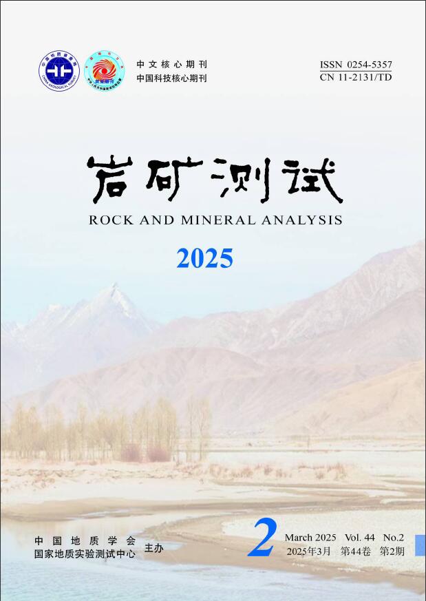
Figure 1.
Morphological characteristics of illite-smectite mixed-layer clay minerals: (a) Three-dimensional stacking of the growth layer; (b) Transverse morphology characteristics of the growth layer; (c) Longitudinal morphology characteristics of the growth layer.
-
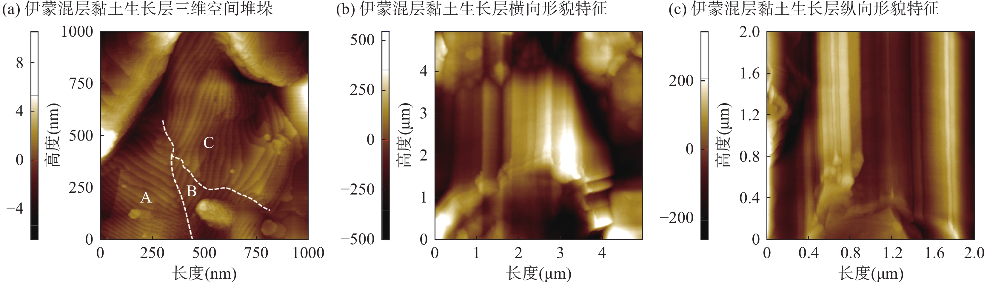
Figure 2.
Morphologic characteristics of chlorite: (a) Chlorite longitudinal no-gap superposition; (b) Plane morphology of chlorite growth layer; (c) Near-regular polygon of chlorite
-
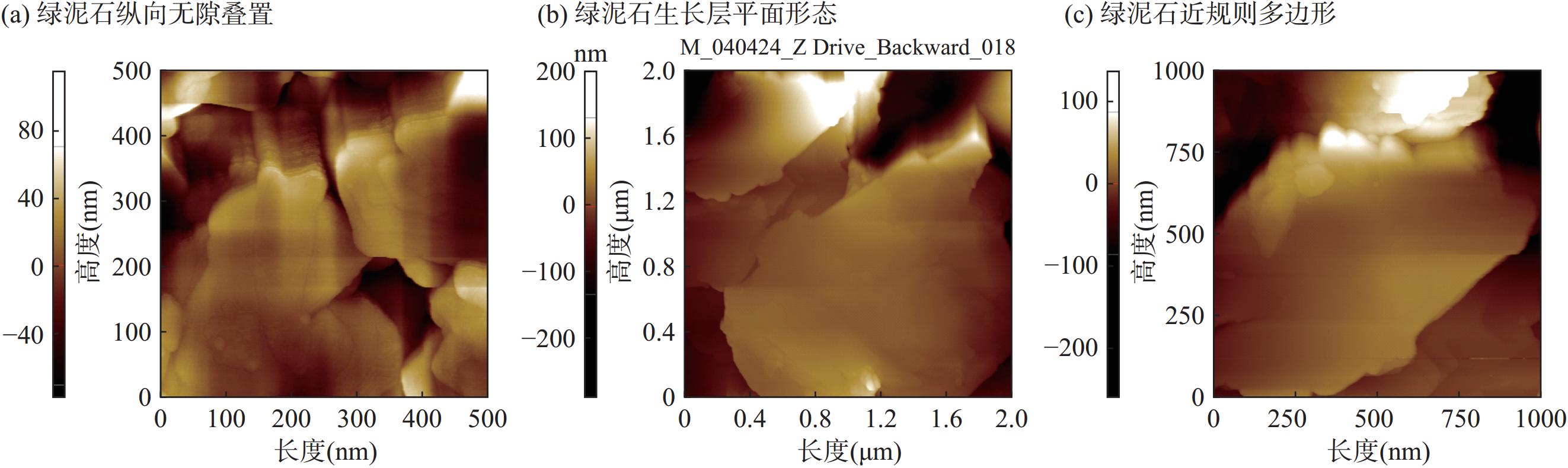
Figure 3.
Mineral morphologies of illite in 10μm×10μm large field of view: (a) Morphology characteristics of illite microcrystalline aggregate; (b) Morphology of scaly illite; (c) Morphological characteristics of illite aggregate.
-
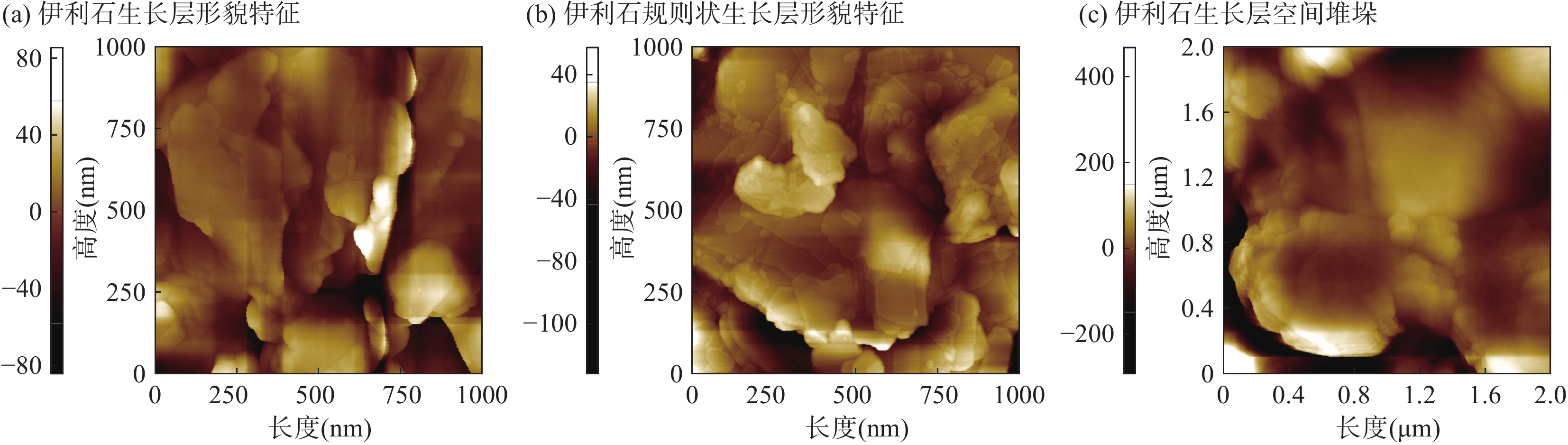
Figure 4.
Mineral morphologies of illite in 1μm×1μm fine scanning images: (a) Morphology of illite growth layer; (b) Morphology of illite regular growth layer; (c) Illite growth layer space stacking.