
| Professional Committee of Rock and Mineral Testing Technology of the Geological Society of China, National Geological Experiment and Testing Center | Host |
| Citation: |
HU Yaoyao, WANG Haozheng, HOU Yuyang, SONG Haoran. A Method for Estimating Micro-area Composition of Quartz-diorite Based on Quantitative Mapping of Electron Probe Microanalysis[J]. Rock and Mineral Analysis, 2022, 41(2): 260-271. doi: 10.15898/j.cnki.11-2131/td.202109280132

|
A Method for Estimating Micro-area Composition of Quartz-diorite Based on Quantitative Mapping of Electron Probe Microanalysis
-
Abstract
BACKGROUND The estimation of bulk composition of the micro-area of a rock is an important basis of tracing rock evolution. The conventional electron probe microanalysis (EPMA) mapping method cannot provide quantitative analysis results of the surface scanning area.
OBJECTIVES In order to estimate the composition of the micro-area by quantitative mapping of EPMA.
METHODS The mapping and point analyses were carried out to determine the composition of the micro-area of a homogeneous quartz-diorite. By performing image correction of the pixels of the mapping of the major elements, and using the point analysis data after ZAF correction and the gray value of the surface scan image to perform the least square curve fitting method, the X-ray intensity and concentration were converted.
RESULTS By comparing to X-ray fluorescence spectrometry (XRF), the relative errors of SiO2, CaO, FeO, Al2O3 and TiO2 of EPMA method were within 10%, and the relative standard deviation was less than 10%. The relative error and standard deviation of MgO and Na2O were slightly larger, which can be improved by multiple measurements. The K2O content were not accurate due to lack of K-rich silicate minerals' point analysis.
CONCLUSIONS The results show that the estimation of micro-area composition of a rock with relatively homogeneous mineral distributions can be carried out by using point analyses of EPMA to correct the mapping under good instrument conditions, and the influence of mineral morphology, particle size, and distribution can be reduced by multiple measurements on different sections.
-

-
References
[1] Wang H Z, Zhang H F, Zhai M G, et al. Granulite facies metamorphism and crust melting in the Huai'an terrane at~1.95Ga, North China Craton: New constraints from geology, zircon U-Pb, Lu-Hf isotope and metamorphic conditions of granulites[J]. Precambrian Research, 2016, 286: 126-151. doi: 10.1016/j.precamres.2016.09.012 [2] 孙春青, 游海涛, 刘嘉麒, 等. 火山灰全岩与原位分析差异: 以四海龙湾记录的1600年前金龙顶子火山喷发为例[J]. 地球科学, 2016, 41(1): 97-104. Sun C Q, You H T, Liu J L, et al. Differences between whole rock and in situ analysis on Tephra: Evidence from the 1600a cal. BP eruption of Jinlongingzi Volcano[J]. Earth Science, 2016, 41(1): 97-104. [3] Wang H Z, Laurent O, Zhang H F, et al. Recognition and significance of c. 800Ma upper amphibolite to granulite facies metamorphism in metasedimentary rocks from the NW margin of the Yangtze Block[J]. Journal of the Geological Society, 2020, 177(2): 424-441. doi: 10.1144/jgs2019-035 [4] 许乃岑, 沈加林, 张静. X射线衍射-X射线荧光光谱-电子探针等分析测试技术在玄武岩矿物鉴定中的应用[J]. 岩矿测试, 2015, 34(1): 75-81. Xu N C, Shen J L, Zhang J. Application of X-ray diffraction, X-ray fluorescence spectrometry and electron microprobe in the identification of basalt[J]. Rock and Mineral Analysis, 2015, 34(1): 75-81. [5] 周伟, 曾梦, 王健, 等. 熔融制样-X射线荧光光谱法测定稀土矿石中的主量元素和稀土元素[J]. 岩矿测试, 2018, 37(3): 298-305. Zhou W, Zeng M, Wang J, et al. Determination of major and rare earth elements in rare earth ores by X-ray fluorescence spectrometry with fusion sample preparation[J]. Rock and Mineral Analysis, 2018, 37(3): 298-305. [6] 李小莉, 吴良英, 王力强, 等. X射线荧光光谱法分析碳酸盐时两种制样方法的比较[J]. 理化检验(化学分册), 2016, 52(6): 664-668. Li X L, Wu L Y, Wang L Q, et al. Comparative study on the 2 methods for preparation of sample discs in XRFs analysis of carbonates[J]. Physical Testing and Chemical Analysis (Part B: Chemical Analysis), 2016, 52(6): 664-668. [7] 汪方跃, 葛粲, 宁思远, 等. 一个新的矿物面扫描分析方法开发和地质学应用[J]. 岩石学报, 2017, 33(11): 3422-3436. Wang F Y, Ge C, Ning S Y, et al. A new approach to LA-ICP-MS mapping and application in geology[J]. Acta Petrologica Sinica, 2017, 33(11): 3422-3436. [8] Spear F S, Wolfe O M. Evaluation of the effective bulk composition (EBC) during growth of garnet[J]. Chemical Geology, 2018, 491: 39-47. doi: 10.1016/j.chemgeo.2018.05.019 [9] 田作林, 张泽明, 董昕. 有效全岩成分的计算方法及其在变质相平衡模拟中的应用[J]. 岩石学报, 2020, 36(9): 2616-2630. Tian Z L, Zhang Z M, Dong X. Calculation of effective bulk composition and its application in metamorphic phase equilibria modeling[J]. Acta Petrologica Sinica, 2020, 36(9): 2616-2630. [10] 聂潇, 王宗起, 陈雷, 等. 蚀变粗面岩中再平衡结构黑云母的电子探针分析[J]. 岩矿测试, 2019, 38(5): 565-574. Nie X, Wang Z Q, Chen L, et al. Electron microprobe analysis of biotite with reequilibration texture in altered trachyte[J]. Rock and Mineral Analysis, 2019, 38(5): 565-574. [11] 秦玉娟, 余朝丰, 徐洋, 等. 应用电子探针原位微区分析技术测试硅硼钠石矿物[J]. 电子显微学报, 2016, 35(3): 217-222. doi: 10.3969/j.issn.1000-6281.2016.03.006 Qin Y J, Yu C F, Xu Y, et al. A microarea analysis technique of EPMA to probe reedmergnerite[J]. Journal of Chinese Electron Microscopy Society, 2016, 35(3): 217-222. doi: 10.3969/j.issn.1000-6281.2016.03.006 [12] Reed S J B. Electron microprobe analysis and scanning electron microscopy in geology[M]. Cambridge: Cambridge University Press, 2005: 164. [13] Batanova V G, Sobolev A V, Magnin V. Trace element analysis by EPMA in geosciences: Detection limit, precision and accuracy[C]//IOP Conference Series: Materials Science and Engineering, 2018, 304. [14] Ritchie N. Embracing uncertainty: Modeling the standard uncertainty in electron probe microanalysis—Part Ⅰ[J]. Microscopy and Microanalysis, 2020, 26(3): 469-483. doi: 10.1017/S1431927620001555 [15] Lanari P, Alice V, Thomas B, et al. Quantitative compositional mapping of mineral phases by electron probe micro-analyser[R]. London: Geological Society (Special Publications), 2019: 39-63. [16] Donovan J, Allaz J, Handt A, et al. Quantitative WDS com-positional mapping using the electron microprobe[J]. The American Mineralogist, 2021, 106: 1717-1735. doi: 10.2138/am-2021-7739 [17] Yasumoto A, Yoshida K, Kuwatani T, et al. A rapid and precise quantitative electron probe chemical mapping technique and its application to an ultrahigh-pressure eclogite from the Moldanubian Zone of the Bohemian Massif (Nové Dvory, Czech Republic)[J]. American Mineralogist, 2018, 103(10): 1690-1698. doi: 10.2138/am-2018-6323CCBY [18] Ortolano G, Visalli R, Godard G, et al. Quantitative X-ray map analyser (Q-XRMA): A new GIS-based statistical approach to mineral image analysis[J]. Computers and Geosciences, 2018, 115: 56-65. doi: 10.1016/j.cageo.2018.03.001 [19] 周剑雄, 毛水和, 陈克樵, 等. 电子探针分析[M]. 北京: 地质出版社, 1988: 499. Zhou J X, Mao S H, Chen K Q, et al. Electron probe micro-analysis[M]. Beijing: Geological Publishing, 1998: 499. [20] 梁细荣, 姚立, 田地. 电子探针背景扣除和谱线干扰修正方法的进展[J]. 岩矿测试, 2008, 27(1): 49-54. doi: 10.3969/j.issn.0254-5357.2008.01.013 Liang X R, Yao L, Tian D. Progress in background subtraction and spectral interference correction in electron probe microanalysis[J]. Rock and Mineral Analysis, 2008, 27(1): 49-54. doi: 10.3969/j.issn.0254-5357.2008.01.013 [21] Newbury D E, Marinenko R B, Myklebust R L, et al. Quantitative compositional mapping with the electron probe microanalyzer[M]. Springer Press, 1991: 335-369. [22] 毕秀丽, 邱雨檬, 肖斌, 等. 基于统计特征的图像直方图均衡化检测方法[J]. 计算机学报, 2021, 44(2): 292-303. Bi X L, Qiu Y M, Xiao B, et al. Histogram equalization detection based on statistical features in digital image[J]. Chinese Journal of Computers, 2021, 44(2): 292-303. [23] Anupama S, Prajwalasimha S N, Swapna H. Image de-noising algorithm based on filtering and histogram equalizationl[C]//Proceedings of the Second International Conference on I-SMAC, 2019: 325-328. [24] 李明辉, 郜鲜辉, 吴金金, 等. 电子探针波谱仪和能谱仪在材料分析中的应用及对比[J]. 电子显微学报, 2020, 39(2): 218-223. doi: 10.3969/j.issn.1000-6281.2020.02.018 Li M H, Gao X H, Wu J J, et al. The application and comparison of EPMA WDS and EDS in material analysis[J]. Journal of Chinese Electron Microscopy Society, 2020, 39(2): 218-223. doi: 10.3969/j.issn.1000-6281.2020.02.018 [25] Joseph D G, Charles H. High count rate electron probe microanalysis[J]. Journal of Research of the National Institute of Standards and Technology, 2002, 107(6): 503-508. doi: 10.6028/jres.107.043 [26] Chen B H, Tseng Y, Yin J L. Gaussian-adaptive bilateral filter[J]. IEEE Signal Processing Letters, 2020, 27: 1670-1674. doi: 10.1109/LSP.2020.3024990 [27] 杨赞伟, 郑亮亮, 曲宏松, 等. 联合均值滤波与泊松核双边滤波降噪算法研究[J]. 计算机仿真, 2020, 37(9): 460-463, 468. Yang Z W, Zheng L L, Qu H S, et al. Image denoising research of combining mean filtering with Poisson Kernel improved bilateral filtering[J]. Computer Simulation, 2020, 37(2): 460-463, 468. [28] Kikongi P, Salvas J, Gosselin R. Curve-fitting regression: Improving light element quantification with XRF[J]. X-Ray Spectrometry, 2017, 46(5): 347-355. doi: 10.1002/xrs.2760 [29] Willis K V, Srogi L A, Lutz T, et al. Phase composition maps integrate mineral compositions with rock textures from the micro-meter to the thin section scale[J]. Computers and Geosciences, 2017, 109: 162-177. doi: 10.1016/j.cageo.2017.08.009 [30] 张迪, 陈意, 毛骞, 等. 电子探针分析技术进展及面临的挑战[J]. 岩石学报, 2019, 35(1): 261-274. Zhang D, Chen Y, Mao Q, et al. Progress and challenge of electron probe microanalysis technique[J]. Acta Petrologica Sinica, 2019, 35(1): 261-274. [31] 李德忍. 电子探针分析过程中钠不稳定机理的新解释初探[J]. 岩矿测试, 1995, 14(3): 224-226. Li D R. An explanation for the unstability of sodium determination in the electron microprobe analysis of Albite[J]. Rock and Mineral Analysis, 1995, 14(3): 224-226. [32] 张文兰, 胡欢, 谢磊, 等. Na元素的EPMA定量分析: 矿物晶体结构对Na行为的制约[J]. 高校地质学报, 2021, 27(3): 327-339. Zhang W L, Hu H, Xie L, et al. Quantitative analysis of Na by EPMA: Constraints for the behavior of Na by the crystal structure[J]. Geological Journal of China Universities, 2021, 27(3): 327-339. [33] 杨宗锋, 罗照华, 黄丹峰, 等. 花岗质岩石中矿物含量和粒径对代表性样品的约束[J]. 北京大学学报(自然科学版), 2010, 46(6): 951-959. Yang Z F, Luo Z H, Huang D F, et al. Constraints on representative samples from crystal contents and size in granitic rocks[J]. Acta Scientiarum Naturalium Universitatis Pekinensis, 2010, 46(6): 951-959. [34] 杨宗锋, 李解, 姜晓杰, 等. 火成岩结构的二维定量化分析方法[J]. 地学前缘, 2020, 27(5): 23-38. Yang Z F, Li J, Jiang X J, et al. Two dimensional quantitative textural analysis method for igneous rock[J]. Earth Science Frontiers, 2020, 27(5): 23-38. [35] Hezel D C. A model for calculating the errors of 2D bulk analysis relative to the true 3D bulk composition of an object, with application to chondrules[J]. Computers and Geosciences, 2007, 33(9): 1162-1175. doi: 10.1016/j.cageo.2007.01.003 [36] Richard M P, Owen M W, David J W, et al. Quantifying geological uncertainty in metamorphic phase equilibria modelling; a Monte Carlo assessment and implications for tectonic interpretations[J]. Geoscience Frontiers, 2016, 7(4): 591-607. doi: 10.1016/j.gsf.2015.08.005 [37] McCanta M C, Dyar M D, Dobosh P A. Extracting bulk rock properties from microscale measurements: Subsampling and analytical guidelines[J]. GSA Today, 2017, 27(7): 4-9. [38] Sharma J, Naik A, Pant N, et al. Estimation of effective bulk composition—Critical appraisal and a scanning electron microscope based approach[J]. Geological Journal, 2021, 56(6): 2950-2962. doi: 10.1002/gj.4077 [39] Hombourger C, Outrequin M. Quantitative analysis and high-resolution X-ray mapping with a field emission electron microprobe[J]. Microscopy Today, 2013, 21(3): 10-15. doi: 10.1017/S1551929513000515 [40] Takahashi H, Mcswiggen P, Nielsen C. A unique wavelength- dispersive soft X-ray emission spectrometer for electron probe X-ray microanalyzers[J]. Microscopy and Analysis, 2014, 15: 5-8. [41] Llovet X, Moy A, Pinard P T, et al. Electron probe microanalysis: A review of recent developments and applications in materials science and engineering[J]. Progress in Materials Science, 2021, 116: 100673. -
Access History
Figures(4)
Tables(4)

Export File
Citation
HU Yaoyao, WANG Haozheng, HOU Yuyang, SONG Haoran. A Method for Estimating Micro-area Composition of Quartz-diorite Based on Quantitative Mapping of Electron Probe Microanalysis[J]. Rock and Mineral Analysis, 2022, 41(2): 260-271. doi: 10.15898/j.cnki.11-2131/td.202109280132
Format
Content
 DownLoad:
-
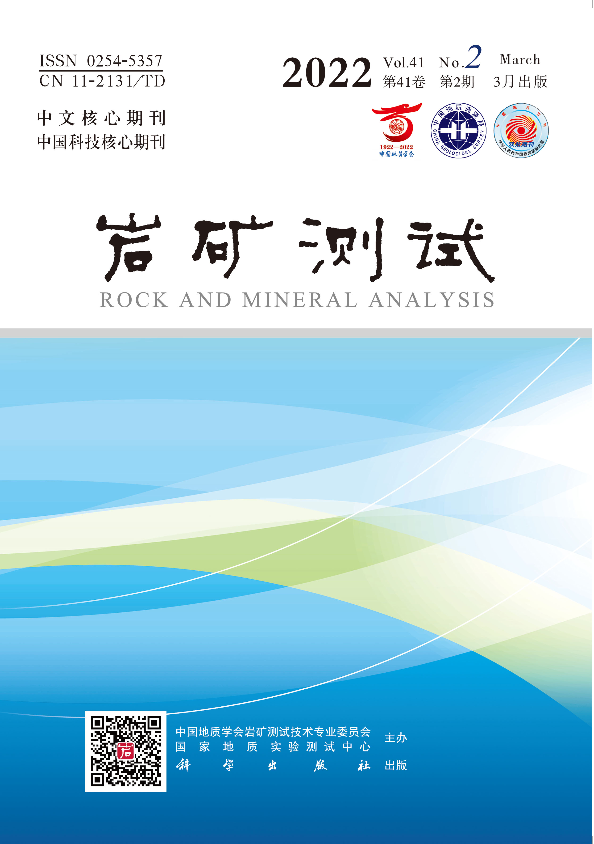
Figure 1.
Field outcrop and microscopic characteristics of Neoproterozoic magmatic quartz-diorite in Micangshan, northern Sichuan. (a, d—Field photos of quartz-diorite which has subhedral granular texture and massive structure; b, c—Plagioclase is enhedral with a turbid surface, and the quartz is subhedral to anhedral distributed around amphibole and plagioclase; e, f—Amphibole is subhedral to anhedral, and contain opaque metallic minerals are founded in amphibole and plagioclase. Mineral abbreviation: Amp—amphibole; Pl—plagioclase; Qz—quartz; Opq—opaque mineral)
-
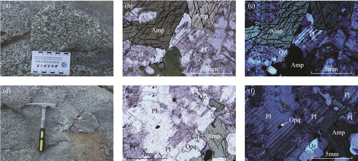
Figure 2.
Backscattered electron image of mapping area and positions of point analyses (Dots represent the position of the simulated point analyses and the numbers in the figure represent the serial number of the article test data. a—The plagioclase formed in the early stage is enhedral crystal. b—The middle part of enhedral plagioclase is alterated into epidote. c—The edge of anhedral amphibole is covered with quartz, ilmenite and K-rich silicate minerals, while apatite, ilmenite and other inclusions exist in it. d—Subhedral ilmenite and magnetite are distributed at the edge of plagioclase and amphibole)
-
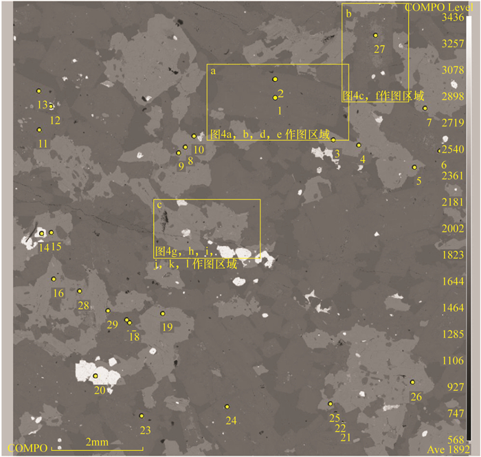
Figure 3.
Curve fitting results of grayscale of pixels and point analyses
-
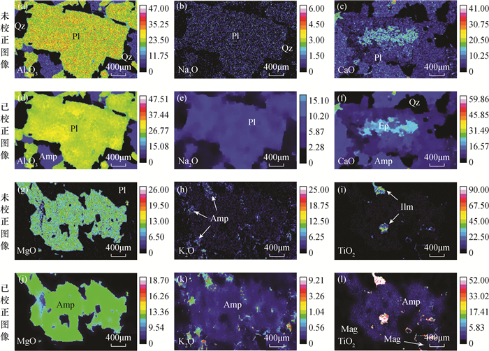
Figure 4.
Phase composition mappings of the minerals. (a, d—The composition of Al2O3 of plagioclase is relatively homogeneous in the corrected image compared to the uncorrected mapping, showing a weak change in modal abundance. b, e— In mapping of Na2O, the composition of plagioclase is inhomogeneous and the count is lost. However, after image processing the composition of plagioclase is homogeneous. c, f— In CaO mapping, the interior of plagioclase is altered. g, j—The composition of minerals is homogeneou and MgO content of amphibole is relatively high in the corrected image compared to the uncorrected mapping, containing ilmenite and other inclusions. h, k—In K2O mapping, amphibole and plagioclase are difficult to distinguish and K-rich silicate minerals exist at the edge. However, after image processing the K2O content of amphibole is obviously higher than that of plagioclase and K-rich silicate minerals are obvious. i, l—Amphibole contains a small amount of TiO2, grain boundary is clear, and ilmenite and magnetite are distributed at the edge in the corrected image compared to the uncorrected mapping. Mineral abbreviation: Amp—amphibole; Pl—plagioclase; Qz—quartz; Ilm—ilmenite; Mag—magnetite)