
| Professional Committee of Rock and Mineral Testing Technology of the Geological Society of China, National Geological Experiment and Testing Center | Host |
| Citation: |
Jing RAN, Chuang-feng GUO, Gu DU, Feng-yu WANG. Quantitative Analysis of Mineral Composition of Kyanite by X-ray Diffraction with Rietveld Refinement Method[J]. Rock and Mineral Analysis, 2019, 38(6): 660-667. doi: 10.15898/j.cnki.11-2131/td.201902220025

|
Quantitative Analysis of Mineral Composition of Kyanite by X-ray Diffraction with Rietveld Refinement Method
-
Abstract
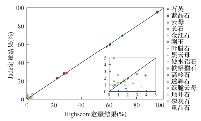
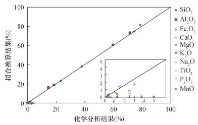
BACKGROUNDThe content of minerals in kyanite can be analyzed by chemical phase method, but the analytical process is very tedious. Moreover, the existence of heteromorphism and refractory minerals can affect the accuracy of analysis results. An internal standard method by X-ray diffraction (XRD) for direct determination of kyanite requires the use of pure mineral to draw a standard curve, but it is extremely challenging to purify minerals due to the inclusion. The differences between the mineral components of kyanite in different mining areas makes the above two methods applicable only to the quantitative analysis of minerals in the same mining area. OBJECTIVESTo simplify analysis the process of mineral composition of kyanite and improve efficiency. METHODSThe content of minerals in kyanite was studied by X-ray diffraction. The X-ray diffraction Rietveld refinement method was used to analyze the secondary standard materials and field samples of kyanite, and the results were compared with the diffraction quantitative method and chemical analysis results including RIR method, adiabatic method and K-value method. RESULTSRietveld refinement method was simple and effective in correcting the diffraction intensity error caused by the preferred orientation. Accurate results can be obtained by Rietveld refinement method more than other methods of X-ray quantitative phase analysis, such as RIR method, adiabatic method, K-value method. The analytic absolute error of minerals with content greater than 5% was less 1%, and significantly lower than the allowable error. The results obtained by Highscore agree well with results measured by Jade, and the double difference of results was less than 0.8%. The relative standard deviation was less than 2.5%. The spiked recoveries of kyanite were 95.3%-101.0%. CONCLUSIONSX-ray diffraction with Rietveld refinement method is simple, and can be used to determine the content of all minerals in kyanite from different mining areas. -
Keywords:
- kyanite /
- phase composition /
- X-ray diffraction /
- Rietveld refinement method /
- preferred orientation
-

-
References
[1] 王梅英.蓝晶石的化学物相分析[J].非金属矿, 2006, 29(1):15-16. Wang M Y.Chemical phase analysis of cyanite[J].Non-Metallic Mines, 2006, 29(1):15-16. [2] 王梅英, 李鹏程, 李艳华, 等.蓝晶石矿中氟钠镁铝硅铁钛钾钙元素的X射线荧光光谱分析[J].岩矿测试, 2013, 32(6):909-914. doi: 10.3969/j.issn.0254-5357.2013.06.011 Wang M Y, Li P C, Li Y H, et al.Analysis of F, Na, Mg, Al, Si, Fe, Ti, K and Ca in cyanite ores by X-ray fluorescence spectrometry[J].Rock and Mineral Analysis, 2013, 32(6):909-914. doi: 10.3969/j.issn.0254-5357.2013.06.011 [3] 杜晓冉, 智红梅, 张金矿, 等.X射线衍射法定量分析蓝晶石样品[J].理化检验(化学分册), 2013, 49(4):402-404. Du X R, Zhi H M, Zhang J K, et al.Quantitative analysis of kyanite sample with X-ray diffractometry[J].Physical Testing and Chemical Analysis (Part B:Chemical Analysis), 2013, 49(4):402-404. [4] 邱贤荣, 齐砚勇, 唐志强.全谱拟合定量分析石灰石[J].分析科学学报, 2013, 29(1):146-148. Qiu X R, Qi Y Y, Tang Z Q.Rietveld quantitative analysis of limestone[J].Journal of Analytical Science, 2013, 29(1):146-148. [5] 冉敬, 杜谷, 王凤玉.X射线衍射全谱拟合法快速分析长石矿物含量[J].岩矿测试, 2017, 36(5):489-494. Ran J, Du G, Wang F Y.Rapid analysis of feldspar by X-ray diffractometry Rietveld refinement method[J].Rock and Mineral Analysis, 2017, 36(5):489-494. [6] 迟广成, 肖刚, 汪寅夫, 等.铁矿石矿物组分的X射线粉晶衍射半定量分析[J].冶金分析, 2015, 35(1):38-44. Chi G C, Xiao G, Wang Y F, et al.Semi-quantitative analysis of the mineral components of iron ores by X-ray powder diffraction[J].Metallurgical Analysis, 2015, 35(1):38-44. [7] 洪汉烈, 陈建军, 杨淑珍, 等.水泥熟料定定量分析的全谱拟合法[J].分析测试学报, 2001, 20(2):5-8. doi: 10.3969/j.issn.1004-4957.2001.02.002 Hong H L, Chen J J, Yang S Z, et al.Quantitative phase analysis of cement clinker by Rietveld full pattern fitting method[J].Journal of Instrumental Analysis, 2001, 20(2):5-8. doi: 10.3969/j.issn.1004-4957.2001.02.002 [8] Gualtieri M L, Romagnoli M, Miselli P, et al.Full quantitative phase analysis of hydrated lime using the Rietveld method[J].Cement & Concrete Research, 2012, 42(9):1273-1279. [9] Santini T C.Application of the Rietveld refinement method for quantification of mineral concentrations in bauxite residues (alumina refining tailings)[J].International Journal of Mineral Processing, 2015, 139:1-10. doi: 10.1016/j.minpro.2015.04.004 [10] Woodruff L, Cannon W F, Smith D B, et al.The distribu-tion of selected elements and minerals in soil of the conterminous United States[J].Journal of Geochemical Exploration, 2015, 154:49-60. doi: 10.1016/j.gexplo.2015.01.006 [11] Gentili S, Comodi P, Bonadiman C, et al.Mass balance vs Rietveld refinement to determine the modal composition of ultramafic rocks:The case study of mantle peridotites from Northern Victoria Land (Antarctica)[J].Tectonophysics, 2015, 650:144-155. doi: 10.1016/j.tecto.2015.01.024 [12] 肖松.河南隐山蓝晶石矿物成因的研究[J].中国矿业, 2005, 14(2):47-49. doi: 10.3969/j.issn.1004-4051.2005.02.015 Xiao S.On mineral genesis of Yinsan kyanite in Henan[J].China Mineral Magazine, 2005, 14(2):47-49. doi: 10.3969/j.issn.1004-4051.2005.02.015 [13] 李锁成, 张松林, 火军昌, 等.萨尔哈布塔勒蓝晶石矿床地质及矿物学特征[J].矿产保护与利用, 2015(1):12-15. Li S C, Zhang S L, Huo J C, et al.The process mineralogy and geological characters of Saerhabutale Kyanite deposit in Gansu Province[J].Conservation and Utilization of Mineral Resources, 2015(1):12-15. [14] 万红波, 廖立兵.膨润土中蒙脱石物相的定量分析[J].硅酸盐学报, 2009, 37(12):2055-2060. doi: 10.3321/j.issn:0454-5648.2009.12.017 Wan H B, Liao L B.Quantitative phase analysis of montmorillonite in bentonite[J].Journal of the Chinese Ceramic Society, 2009, 37(12):2055-2060. doi: 10.3321/j.issn:0454-5648.2009.12.017 [15] 房俊卓, 张霞, 徐崇福.实验条件对X射线衍射物相定量分析结果的影响[J].岩矿测试, 2008, 27(1):60-62. doi: 10.3969/j.issn.0254-5357.2008.01.015 Fang J Z, Zhang X, Xu C F.Effect of experimental conditions on X-ray diffractometric quantitative phase analysis[J].Rock and Mineral Analysis, 2008, 27(1):60-62. doi: 10.3969/j.issn.0254-5357.2008.01.015 [16] 马礼敦.X射线粉末衍射的新起点——Rietveld全谱拟合[J].物理学进展, 1996, 16(2):251-271. doi: 10.3321/j.issn:1000-0542.1996.02.005 Ma L D.The new starting point of X-ray powder diffraction-Rietveld whole pattern fitting[J].Progress in Physics, 1996, 16(2):251-271. doi: 10.3321/j.issn:1000-0542.1996.02.005 [17] 林伟伟, 宋友桂.沉积物中X射线衍射物相定量分析中的两种方法对比研究[J].地球环境学报, 2017, 8(1):83-86. Lin W W, Song Y G.A comparative study on X-ray diffraction mineral quantitative analysis of two methods in sediments[J].Journal of Earth Enviroment, 2017, 8(1):83-86. [18] 伍月, 刘欣, 张波, 等.X射线粉晶衍射基体清洗法在矿物定量分析中的应用[J].地质与资源, 2017, 26(3):323-328. Wu Y, Liu X, Zhang B, et al.The application and research of X-ray powder diffraction matrix flushing method in quantitative analysis[J].Geology and Resources, 2017, 26(3):323-328. [19] 施倪承, 白文吉, 马喆生, 等.西藏罗布莎铬铁矿床中的金刚石包体X射线衍射研究[J].地质学报, 2002, 76(4):496-500. doi: 10.3321/j.issn:0001-5717.2002.04.008 Shi N C, Bai W J, Ma Z S, et al.A study of X-ray diffraction of diamond inclusions from Luobusha, Tibet[J].Acta Geologica Sinica, 2002, 76(4):496-500. doi: 10.3321/j.issn:0001-5717.2002.04.008 [20] 王德强, 王辅亚, 王冠鑫, 等.钾含量对白云母X射线衍射特征的影响[J].大地构造与成矿学, 2004, 28(1):69-73. doi: 10.3969/j.issn.1001-1552.2004.01.010 Wang D Q, Wang F Y, Wang G X, et al.XRD character of muscovites with different potassium contents[J].Geotectonica et Metallogenia, 2004, 28(1):69-73. doi: 10.3969/j.issn.1001-1552.2004.01.010 [21] 杜谷, 王坤阳, 冉敬, 等.红外光谱/扫描电镜等现代大型仪器岩石矿物鉴定技术及其应用[J].岩矿测试, 2014, 33(5):625-633. doi: 10.3969/j.issn.0254-5357.2014.05.003 Du G, Wang K Y, Ran J, et al.Application of IR/SEM and other modern instruments for mineral identification[J].Rock and Mineral Analysis, 2014, 33(5):625-633. doi: 10.3969/j.issn.0254-5357.2014.05.003 -
Access History
Figures(5)
Tables(4)

Export File
Citation
Jing RAN, Chuang-feng GUO, Gu DU, Feng-yu WANG. Quantitative Analysis of Mineral Composition of Kyanite by X-ray Diffraction with Rietveld Refinement Method[J]. Rock and Mineral Analysis, 2019, 38(6): 660-667. doi: 10.15898/j.cnki.11-2131/td.201902220025
Format
Content
 DownLoad:
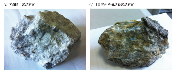
- Figure 1. Rock samples of kyanite from Henan and Gansu Provinces
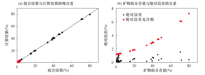
- Figure 2. Comparison of standard addition experiment
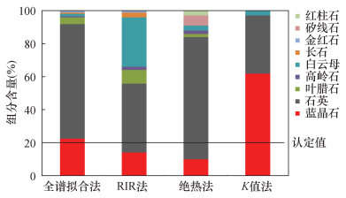
- Figure 3. Comparison of analytical results determined by different X-ray diffraction quantitative methods
- Figure 4. Correlation of analytical results obtained by Highscore and Jade softwares
- Figure 5. Comparison of the chemical components of samples