
| Professional Committee of Rock and Mineral Testing Technology of the Geological Society of China, National Geological Experiment and Testing Center | Host |

| Citation: |
Hai-yan YU, Qing-feng RUAN, Xin SHA, Yu-fu YANG. Study on Color-causing Elements in Qinghai Nephrite by Elemental Analysis and Electron Paramagnetic Resonance Spectroscopy[J]. Rock and Mineral Analysis, 2019, 38(3): 288-296. doi: 10.15898/j.cnki.11-2131/td.201805140130

|
Study on Color-causing Elements in Qinghai Nephrite by Elemental Analysis and Electron Paramagnetic Resonance Spectroscopy
-
Abstract
BACKGROUNDColor is the important manifestation of nephrite value. Qinghai nephrite has different colors, but there is a lack of research on coloration. In recent years, the study on the coloration of nephrite in Qinghai mainly focused on azure-green and blue-violet, and it was considered that Cr3+ and Mn2+ were respective coloration elements of azure-green and blue-violet. The color of Qinghai nephrite is not a simple color, such as white-green, azure-green, and blue-violet. Therefore, Qinghai nephrite should contain a variety of color-causing elements. OBJECTIVESTo reveal the color-causing elements of eight colors of nephrite from Qinghai based on the relationship between analysis data and hue changes. METHODSX-ray Fluorescence Spectrometry (XRF), Chemical Titration, Inductively Coupled Plasma-Mass Spectrometry (ICP-MS) and Electron Paramagnetic Resonance Spectroscopy (EPR) were used to study the color-causing elements of nephrite. RESULTSThe color-causing element of white Qinghai nephrite was Fe3+. The color-causing elements of white-green and azure-green Qinghai nephrite were Fe2+ and Fe3+, respectively. The chromogenic elements of green Qinghai nephrite were Fe2+, Fe3+ and Mn in high valence. The color-causing elements of azure-green Qinghai nephrite were Fe2+, Fe3+ and Cr3+. The chromogenic elements of yellow and brown Qinghai nephrite were Fe3+ and high-valence Mn. Fe3+ and Ti4+ were the color-causing elements of blue-purple Qinghai nephrite. CONCLUSIONSThis study determined the color-causing elements of different colors of Qinghai nephrite, which provided a theoretical basis for the study of the coloration mechanism of Qinghai nephrite. -
Keywords:
- Qinghai nephrite /
- Electron Paramagnetic Resonance /
- color-causing elements /
- Fe
-

-
References
[1] 李雯雯, 吴瑞华.和田玉的颜色及其色度学研究[J].矿物岩石地球化学通报, 1999, 18(4):418-422. Li W W, Wu R H.The colorimetry and chromaticity study of Xinjiang Hetian jade[J].Bulletin of Mineralogy, Petrology and Geochemistry, 1999, 18(4):418-422. [2] 那宝成, 冷莹莹, 李祥虎.软玉致色元素的研究[J].超硬材料工程, 2008, 20(3):55-58. doi: 10.3969/j.issn.1673-1433.2008.03.014 Na B C, Leng Y Y, Li X H.Research on coloring element of nephrite[J].Uperhard Material Engineering, 2008, 20(3):55-58. doi: 10.3969/j.issn.1673-1433.2008.03.014 [3] Wang Y Y, Gan F X.Coloration mechanism and chromaticity of Xiuyan jade of China[J].Spectroscopy and Spectral Analysis, 2012, 32(9):2305-2310. [4] 冯晓燕, 陆太进, 张辉, 等.拉曼光谱分析在软玉颜色评价中的应用[J].矿物岩石, 2015, 35(1):1-6. doi: 10.3969/j.issn.1007-2802.2015.01.001 Feng X Y, Lu T J, Zhang H, et al.Application of Raman spectroscopic technique to investigation on nephrite color[J].Journal of Mineralogy and Petrology, 2015, 35(1):1-6. doi: 10.3969/j.issn.1007-2802.2015.01.001 [5] 韩文, 洪汉烈, 吴钰, 等.和田玉糖玉的致色机理研究[J].光谱学与光谱分析, 2013, 33(6):1446-1450. doi: 10.3964/j.issn.1000-0593(2013)06-1446-05 Han W, Hong H L, Wu Y, et al.Color genesis of brown jade from Hetian nephrite[J].Spectroscopy and Spectral Analysis, 2013, 33(6):1446-1450. doi: 10.3964/j.issn.1000-0593(2013)06-1446-05 [6] 周征宇, 陈盈, 廖宗廷, 等.溧阳软玉的岩石矿物学研究[J].岩石矿物学杂志, 2009, 28(5):490-494. doi: 10.3969/j.issn.1000-6524.2009.05.010 Zhou Z Y, Chen Y, Liao Z T, et al.A petrological and mineralogical study of Liyang nephrite[J].Acta Petrologica et Mineralogica, 2009, 28(5):490-494. doi: 10.3969/j.issn.1000-6524.2009.05.010 [7] 卢保奇.四川石棉软玉猫眼和蛇纹石猫眼的宝石矿物学及其谱学研究[D].上海: 上海大学, 2005. http://cdmd.cnki.com.cn/Article/CDMD-11903-2006176062.htm Lu B Q.The Gemological Mineralogy and Spectroscopy of Nephrite Cat's Eye and Serpentine Cat's Eye from Shimian, Sichuan Province, Southwest of China[D]: Shanghai: Shanghai University, 2005.[8] 杨林.贵州罗甸玉矿物岩石学特征及成因机理研究[D].成都: 成都理工大学, 2013. http://cdmd.cnki.com.cn/Article/CDMD-10616-1013288435.htm Yang L.Study on Petro-mineral Features and Genetic Mechanism of Luodian Jade, Guizhou Province[D].Chengdu: Chengdu University of Technology, 2013.[9] 刘虹靓, 杨明星, 杨天翔, 等.青海翠青玉的宝石学特征及颜色研究[J].宝石和宝石学杂志, 2013, 15(1):7-14. doi: 10.3969/j.issn.1008-214X.2013.01.002 Liu H L, Yang M X, Yang T X, et al.Study on colour and gemological characteristics of viridis nephrite from Qinghai Province[J].Journal of Gems & Gemmology, 2013, 15(1):7-14. doi: 10.3969/j.issn.1008-214X.2013.01.002 [10] 罗泽敏, 沈锡田, 杨明星.青海三岔河灰紫色软玉颜色定量表达与紫色成因研究[J].光谱学与光谱分析, 2017, 37(3):822-828. Luo Z M, Shen X T, Yang M X.Study on color quantitative expression, replication and color origin of gray-purple nephrite from Qinghai, China based on spectroscopy methods[J].Pectroscopy and Spectral Analysis, 2017, 37(3):822-828. [11] Manoogian A.The electron spin resonance of Mn2+ in tremolite[J].Canadian Journal of Physics, 2011, 46(2):129-133. [12] Manoogian A.Intensity of allowed and forbidden electron spin resonance lines of Mn2+ in tremolite[J].Canadian Journal of Physics, 2011, 46(9):1029-1033. [13] Mcgavin D G, Palmer R A, Tennant W C, et al.Use of ultrasonically modulated electron resonance to study S-state ions in mineral crystals:Mn2+, Fe3+in tremolite[J].Physics & Chemistry of Minerals, 1982, 8(5):200-205. [14] Vinokurov V M, Zaripov M M, Stepanov V G.Paramagnetic resonance of Mn2+ions in diopside[J].Solid State Communications, 1964, 2(5):Ⅲ-Ⅳ. [15] Pan Y, Nilges M J.Electron paramagnetic resonance spectroscopy:Basic principles, experimental techniques and applications to earth and planetary sciences[J].Reviews in Mineralogy & Geochemistry, 2014, 78(1):655-690. [16] Vasyukov V N, Shapovalov V V, Schwarz S A, et al.Temperature-induced changes in the EPR spectrum of the magnetic center in kaolin[J].Journal of Magnetic Resonance, 2002, 154(1):15-21. [17] Pan Y, Mao M, Lin J.Single-crystal EPR study of Fe3+ and VO2+ in prehnite from the Jeffrey mine, Asbestos, Quebec[J].Canadian Mineralogist, 2009, 47(4):933-945. doi: 10.3749/canmin.47.4.933 [18] Balan E.Structural Fe3+ in natural kaolinites:New insights from electron paramagnetic resonance spectra fitting at X and Q-band frequencies[J].Clays & Clay Minerals, 1999, 47(5):605-616. [19] Gaite J M, Izotov V V, Nikitin S I, et al.EPR and optical spectroscopy of impurities in two synthetic beryls[J].Applied Magnetic Resonance, 2001, 20(3):307-315. doi: 10.1007/BF03162283 [20] Ollier N, Fuchs Y, Cavani O, et al.Influence of impurities on Cr3+ luminescence properties in Brazilian emerald and alexandrite[J].European Journal of Mineralogy, 2015, 27(6):783-792. doi: 10.1127/ejm/2015/0027-2484 [21] Zheng W C, Zhou Q, Wu X X, et al.Theoretical investigations of the EPR parameters of Ti3+ in beryl crystal[J].Zeitschrift Für Naturforschung A, 2006, 61(5-6):286-288. [22] Yuan Z, Wu X X, Lü H, et al.EPR parameters and defect structures of the off-center Ti3+ ion on the Sr2+ site in neutron-irradiated SrTiO3 crystal[J].Journal of Physics & Chemistry of Solids, 2007, 68(9):1652-1655. [23] 赵敏光.晶体场和电子顺磁共振理论[M].北京:科学出版社, 1991:204-216. Zhao M G.Theory on Crystal Field and Electron Paramagnetic Resonance[M].Beijing:Science Press, 1991:204-216 -
Access History
Figures(5)
Tables(2)

Export File
Citation
Hai-yan YU, Qing-feng RUAN, Xin SHA, Yu-fu YANG. Study on Color-causing Elements in Qinghai Nephrite by Elemental Analysis and Electron Paramagnetic Resonance Spectroscopy[J]. Rock and Mineral Analysis, 2019, 38(3): 288-296. doi: 10.15898/j.cnki.11-2131/td.201805140130
Format
Content
 DownLoad:


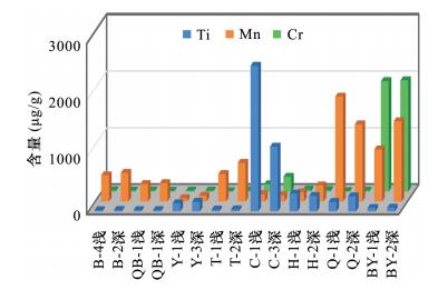
- Figure 1. Histogram of transition metallic elements content in Qinghai nephrites with different colors
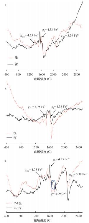
- Figure 2. Comparison of electron paramagnetic resonance spectra in (a) 3010-4010G and (b) 260-6760G in Qinghai nephrites with different colors at room temperature
- Figure 3. Comparison charts of electron paramagnetic resonance in 400-2500G of Fe3+ in Qinghai nephrites with different shades at room temperature
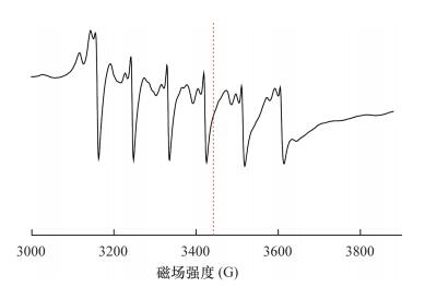
- Figure 4. Electron paramagnetic resonance chart in 3010-4010G of Ti3+ in Qinghai nephrites with different colors at liquid helium temperature
- Figure 5. Comparison charts of electron paramagnetic resonance in 3010-4010G of Mn2+ in Qinghai nephrites with different shades at room temperature