
| Zhengzhou Institute of Multipurpose Utilization of Mineral Resources, Chinese Academy of Geological Sciences | Host |
| Citation: |
WANG Jiali, WANG Jieliang, SHI Jingyang, WU Xu, CAO Zhao. Research Progress on Surface Property Testing Methods for Mineral Flotation[J]. Conservation and Utilization of Mineral Resources, 2025, 45(1): 101-113. doi: 10.13779/j.cnki.issn1001-0076.2025.01.004

|
Research Progress on Surface Property Testing Methods for Mineral Flotation
-
Abstract
Flotation is a technology that separates and purifies materials in a three−phase flow of gas, liquid, and solid based on the differences in physical and chemical properties (mainly referring to wettability) of the material surface. It is widely used for mineral separation. Studying the basic flotation behavior, wettability, surface electrical properties, adsorption, and solution chemistry of minerals is a fundamental method for determining the interaction mechanism between flotation agents and mineral surfaces. However, for many complex flotation systems, various modern testing methods are required to characterize or prove these interaction mechanisms, and to reveal the essence of the interaction between flotation agents and mineral surfaces more clearly at the microscopic level. This article comprehensively analyzes the application and research status of imaging analysis techniques such as atomic force microscopy (AFM), transmission electron microscopy (TEM), scanning electron microscopy (SEM), and surface composition analysis techniques such as Zeta potential, infrared spectroscopy, Raman spectroscopy, X−ray photoelectron spectroscopy (XPS), time of flight secondary ion mass spectrometry (TOF−SIMS) in flotation, providing reference for the better development of flotation interface testing in the future.
-
Keywords:
- flotation /
- test technology /
- imaging analysis /
- surface composition analysis
-

-
References
[1] FILIPPOV L O, SEVEROV V V, FILIPPOVA I V. An overview of the beneficiation of iron ores via reverse cationic flotation[J]. International Journal of Mineral Processing, 2014, 127: 62−69. [2] 赵春花. 原子力显微镜的基本原理及应用[J]. 化学教育(中英文), 2019(4): 10−15. ZHAO C H. Basic principles and applications of atomic force microscopy[J]. Chemistry Education (Chinese and English), 2019(4): 10−15. [3] LI L L L, ZHANG C Z C, YUAN Z Y Z, et al. AFM and DFT study of depression of hematite in oleate−starch−hematite flotation system[J]. Applied Surface Science, 2019(1): 749−758. [4] DONG L, WEI Q, QIN W, et al. Selective adsorption of sodium polyacrylate on calcite surface: Implications for flotation separation of apatite from calcite[J]. Separation and Purification Technology, 2020, 241. [5] DONG L D L, JIAO F J F, QIN W Q W, et al. Activation effect of lead ions on scheelite flotation: adsorption mechanism, AFM imaging and adsorption model[J]. Separation and Purification Technology, 2019, 209: 955−963. doi: 10.1016/j.seppur.2018.09.051 [6] JIN S J S, SHI Q S Q, LI Q L Q, et al. Effect of calcium ionic concentrations on the adsorption of carboxymethyl cellulose onto talc surface: Flotation, adsorption and AFM imaging study[J]. Powder Technology, 2018, 331: 155−161. doi: 10.1016/j.powtec.2018.03.014 [7] BAI T A, YANG F, WANG H, et al. Adhesion forces of shale oil droplet on mica surface with different roughness: An experimental investigation using atomic force microscopy[J]. Energies, 2022, 15(17): 6460. doi: 10.3390/en15176460 [8] 郭姚, 任嗣利. 原子力显微镜技术在矿物浮选中的应用现状[J]. 有色金属(选矿部分), 2022(1): 73−79. GUO Y, REN S L. Application status of atomic force microscopy technology in mineral flotation[J]. Nonferrous Metals (Mineral Processing Part), 2022(1): 73−79. [9] LI H, LONG J, XU Z, et al. Novel polymer aids for low−grade oil sand ore processing.[J]. Canadian Journal of Chemical Engineering, 2009(2): 168−176. [10] YANG J Y, QI S, SONG B, et al. The interaction force between scheelite and scheelite/fluorite/calcite measured using atomic force microscopy[J]. Journal of Chemistry, 2020.https://doi.org/10.1155/2020/3163415. [11] SUN X, LIU W, ZHUO Q, et al. Probing the interaction between coal particle and collector using atomic force microscope and density functional theory calculation[J]. Colloids & Surfaces A: Physicochemical & Engineering Aspects, 2023, 660: 130916. [12] 廖立兵, 王丽娟, 尹京武等. 矿物材料现代测试技术[M]. 北京化学工业出版社, 2010: 259. LIAO L B, WANG L J, YIN J W, et al. Modern testing technology for mineral materials[M]. Beijing Chemical Industry Press, 2010: 259. [13] 李金华, 潘永信. 透射电子显微镜在地球科学研究中的应用[J]. 中国科学(地球科学), 2015(9): 1359−1382. LI J H, PAN Y X. Application of transmission electron microscopy in earth science research[J]. Chinese Science (Earth Science), 2015(9): 1359−1382. [14] 叶美芳, 刘三, 解古巍, 等. 应用扫描电镜−X射线衍射−电子探针研究北山斑岩铜矿区绢英岩中白色云母的特征[J]. 岩矿测试, 2016(2): 166−177. YE M F, LIU S, Jie G W, et al. Study on the characteristics of white mica in sericite in Beishan porphyry copper deposit area using scanning electron microscopy X−ray diffraction electron probe[J]. Rock and Mineral Testing, 2016(2): 166−177. [15] 丁秀云, 马啸华, 王淑英, 等. 纳米级铁氧磁流体磁性粒子的制备工艺及其性能测试[J]. 信阳师范学院学报(自然科学版), 2005(4): 452−453. DING X Y, MA X H, WANG S Y, et al. Preparation process and performance testing of nanoscale ferrite magnetic fluid magnetic particles[J]. Journal of Xinyang Normal University (Natural Science Edition), 2005(4): 452−453. [16] JIAN W, MAO J, LEHMANN B, et al. Lingbaoite, AgTe3, a new silver telluride from the Xiaoqinling gold district, central China[J]. American Mineralogist, 2020(5): 745−755. [17] HE H, YANG Y, MA L, et al. Evidence for a two−stage particle attachment mechanism for phyllosilicate crystallization in geological processes[J]. American Mineralogist, 2021(6): 983−993. [18] GAO WG, CIOBANU, COOK N, et al. Nanoscale study of lamellar exsolutions in clinopyroxene from olivine gabbro: Recording crystallization sequences in iron−rich layered intrusions[J]. American Mineralogist, 2019(2): 244−261. [19] JOHNSON, MURAYAMA, KUSEL K, et al. Polycrystallinity of green rust minerals and their synthetic analogs: Implications for particle formation and reactivity in complex systems[J]. American Mineralogist, 2015(10): 2091−2105. [20] CIOBANU C L, COOK N J, SLATTERY A D, et al. Nanoscale intergrowths in the bastnäsite–synchysite series record transition toward thermodynamic equilibrium[J]. MRS Bulletin, 2022(3): 250−257. [21] 于洪, 王登红, 李文渊, 等. 透射电子显微镜在关键矿产成因机制研究中的应用进展[J]. 矿床地质, 2023(3): 565−578. YU H, WANG D H, LI W Y, et al. Progress in the application of transmission electron microscopy in the study of key mineral genesis mechanisms[J]. Mineral Geology, 2023(3): 565−578. [22] 陈莉, 徐军, 陈晶. 扫描电子显微镜显微分析技术在地球科学中的应用[J]. 中国科学(地球科学), 2015(9): 1347−1358. CHEN L, XU J, CHEN J. Application of scanning electron microscopy microscopic analysis technology in earth science[J]. Chinese Science (Earth Science), 2015(9): 1347−1358. [23] YANG B Y B, WANG D W D, WANG T W T, et al. Effect of Cu2+ and Fe3+ on the depression of molybdenite in flotation[J]. Minerals Engineering, 2019, 130: 101−109. doi: 10.1016/j.mineng.2018.10.012 [24] CHENG K, WU X, TANG H, et al. The flotation of fine hematite by selective flocculation using sodium polyacrylate[J]. Minerals Engineering, 2022, 176. [25] 温利刚, 贾木欣, 王清, 等. 基于扫描电子显微镜的自动矿物学新技术−BPMA及其应用前景[J]. 有色金属(选矿部分), 2021(2): 12−23. WEN L G, JIA M X, WANG Q, et al. A new automatic mineralogy technology based on scanning electron microscopy − BPMA and its application prospects[J]. Nonferrous Metals (Mineral Processing Section), 2021(2): 12−23. [26] 曹玉璐, 曾宇轲, 张元元. 基于扫描电子显微镜的重矿物物源分析方法对比[J]. 现代地质, 2023(2): 475−485. CAO Y L, ZENG Y K, ZHANG Y Y. Comparison of heavy mineral source analysis methods based on scanning electron microscopy[J]. Modern Geology, 2023(2): 475−485. [27] LIU M, CHEN D, HU B, et al. New insights into the activation mechanism of ammonium ions on the malachite sulfidization flotation[J]. Minerals Engineering, 2024: 205. [28] KAMBLE S, AGRAWAL S, CHERUMUKKIL S, et al. Revisiting Zeta potential, the key feature of interfacial phenomena, with applications and recent advancements[J]. Chemistry Select, 2022, 7(1): 1−40. [29] FENG Q F Q, WEN S W S, ZHAO W Z W, et al. Effect of calcium ions on adsorption of sodium oleate onto cassiterite and quartz surfaces and implications for their flotation separation[J]. Separation and Purification Technology, 2018, 200: 300−306. doi: 10.1016/j.seppur.2018.02.048 [30] ZHAO Q Z Q, LIU W L W, WEI D W D, et al. Effect of copper ions on the flotation separation of chalcopyrite and molybdenite using sodium sulfide as a depressant[J]. Minerals Engineering, 2018, 115: 44−52. doi: 10.1016/j.mineng.2017.10.008 [31] TIAN M T M, LIU R L R, GAO Z G Z, et al. Activation mechanism of Fe(Ⅲ) ions in cassiterite flotation with benzohydroxamic acid collector[J]. Minerals Engineering, 2018(1): 31−37. [32] SHI Q S Q, FENG Q F Q, ZHANG G Z G, et al. Electrokinetic properties of smithsonite and its floatability with anionic collector[J]. Colloids & Surfaces A: Physicochemical & Engineering Aspects, 2012, 410, 178−183. [33] JORDENS A, MARION C, KUZMINA O, et al. Surface chemistry considerations in the flotation of bastnasite[J]. Minerals Engineering, 2014, 66/67/68: 119−129. [34] JORDENS A, MARION C, KUZMINA O, et al. Physicochemical aspects of allanite flotation[J]. Journal of Rare Earths, 2014(5): 476−486. [35] TIAN M, ZHANG C, HAN H, et al. Novel insights into adsorption mechanism of benzohydroxamic acid on lead (Ⅱ) activated cassiterite surface: An integrated experimental and computational study[J]. Minerals Engineering, 2018(1): 327−338. [36] HAN H H H, HU Y H Y, SUN W S W, et al. Novel catalysis catalysis mechanisms of benzohydroxamic acid adsorption by lead ions and changes in the−surface of scheelite particles[J]. Minerals Engineering, 2018, 119: 11−22. doi: 10.1016/j.mineng.2018.01.005 [37] QUAST K K Q U. Literature review on the interaction of oleate with non−sulphide minerals using zeta potential[J]. Minerals Engineering, 2016(3): 10−20. [38] QUAST K. The use of zeta potential to investigate the interaction of oleate on hematite(Article)[J]. Minerals Engineering, 2016(1): 130−137. [39] KUBETZKA A, BODE M, PIETZSCH O. Spin−polarized scanning tunneling microscopy with antiferromagnetic probe tips[J]. Physical Review Letters, 2002, 88(5): 057201. [40] TERNES M, LUTZ C P, HIRJIBEHEDIN C F, et al. The force needed to move an atom on a surface[J]. Science, 2008(5866): 1066−1069. [41] 孙鑫, 黄凌云, 胡博, 等. 新型羟肟酸捕收剂的合成及其对孔雀石的捕收机理研究[J]. 矿产保护与利用, 2022(1): 52−60. SUN X, HUANG L Y, HU B, et al. Synthesis of a novel hydroxamic acid collector and its mechanism for capturing malachite[J]. Mineral Protection and Utilization, 2022(1): 52−60. [42] LINH T, CUBA−CHIEM, HUYNH L, RALSTON J, et al. In situ particle film ATR−FTIR studies of CMC adsorption on talc: The effect of ionic strength and multivalent metal ions[J]. Minerals Engineering, 2008(12−14): 1013−1019. [43] 许鹏云, 李晶, 陈洲, 等. 红外光谱分析技术在浮选过程中的应用研究进展[J]. 光谱学与光谱分析, 2017(8): 2389−2396. XU P Y, LI J, CHEN Z, et al. Research progress on the application of infrared spectroscopy analysis technology in flotation process[J]. Spectroscopy and Spectral Analysis, 2017(8): 2389−2396. [44] 饶金山, 何晓娟, 罗传胜, 等. 辛基羟肟酸浮选氟碳铈矿机制研究[J]. 中国稀土学报, 2015(3): 370−377. RAO J S, HE X J, LUO C S, et al. Study on the mechanism of octylhydroxamic acid flotation of fluorocarbon cerium ore[J]. Chinese Journal of Rare Earth Sciences, 2015(3): 370−377. [45] YIN W, SUN H, HONG J, et al. Effect of Ca selective chelator BAPTA as depressant on flotation separation of magnesite from dolomite[J]. Minerals Engineering, 2019: 144. [46] YANG B, YIN W, ZHU Z. Differential adsorption of hydrolytic polymaleic anhydride as an eco−friendly depressant for the selective flotation of apatite from dolomite[J]. Separation and Purification Technology, 2021(256): 117803. [47] 周枫然, 韩桥, 张体强, 等. 傅里叶变换红外光谱技术的应用及进展[J]. 化学试剂, 2021(8): 1001−1009. ZHOU F R, HAN Q, ZHANG T Q, et al. Application and progress of Fourier transform infrared spectroscopy technology[J]. Chemical Reagents, 2021(8): 1001−1009. [48] 叶军建, 张覃, 侯波, 等. 显微−反射傅里叶变换红外光谱研究油酸钠与胶磷矿吸附机理[J]. 光谱学与光谱分析, 2018(10): 3036−3040. YE J J, ZHANG Q, HOU B, et al. Study on the adsorption mechanism of sodium oleate and collophane by micro reflection Fourier transform infrared spectroscopy[J]. Spectroscopy and Spectral Analysis, 2018(10): 3036−3040. [49] CHENG Q, MEI G, XU W, et al. Flotation of quartz using imidazole ionic liquid collectors with different counterions[J]. Minerals Engineering, 2022: 180. [50] SAHOO H, RATH S S, DAS B, et al. Flotation of quartz using ionic liquid collectors with different functional groups and varying chain lengths[J]. Minerals Engineering, 2016: 107−112. [51] WANG X, LIU W, LIU W, et al. Understanding adsorption of amine surfactants on the solvated quartz (101) surface by a jointed Dreiding−ClayFF force field[J]. Applied Surface Science, 2021, 566. [52] YANG K, KAS R, SMITH W A. In situ infrared spectroscopy reveals persistent alkalinity near electrode surfaces during CO2 electroreduction[J]. Journal of the American Chemical Society, 2019(40): 15891−15900. [53] MENG J, XU L, WANG D, et al. The activation mechanism of metal ions on spodumene flotation from the perspective of in situ ATR−FTIR and ToF−SIMS[J]. Minerals Engineering, 2022: 182. [54] GAO J, SUN W, LYU F. Understanding the activation mechanism of Ca2+ ion in sodium oleate flotation of spodumene: A new perspective[J]. Chemical Engineering Science, 2021(244): 116742. [55] LUO L, WU H, XU L, et al. An in situ ATR−FTIR study of mixed collectors BHA/DDA adsorption in ilmenite−titanaugite flotation system[J]. International Journal of Mining Science and Technology, 2021(4): 689−697. [56] FREDRIKSSON A, LARSSON M L, HOLMGREN A. N−Heptyl xanthate adsorption on a ZnS layer synthesized on germanium: An in situ attenuated total reflection IR study[J]. Journal of Colloid and Interface Science, 2005(1): 1−6. [57] SE A F A F, HOLMGREN A, FORSLING W. Kinetics of collector adsorption on mineral surfaces[J]. Minerals Engineering, 2006(6/7/8): 784−789. [58] 李小军, 卢雪, 任宏江, 等. HmTiSin(m=1~2; n=2~8)团簇结构、电子性质和红外光谱的密度泛函理论研究[J]. 光谱学与光谱分析, 2019(1): 65−72. LI X J, LU X, REN H J, et al. HmTiSin(m=1~2; density functional theory study oncluster structure, electronic properties, and infrared spectroscopy (n=2~8)[J]. Spectroscopy and Spectral Analysis, 2019 (1): 65−72. [59] WANHALA A K, DOUGHTY B, BRYANTSEV V S, et al. Adsorption mechanism of alkyl hydroxamic acid onto bastnäsite: Fundamental steps toward rational collector design for rare earth elements[J]. Journal of Colloid and Interface Science, 2019, 553: 210−219. doi: 10.1016/j.jcis.2019.06.025 [60] 张曦, 南瑞华, 坚佳莹, 等. 拉曼光谱技术及其应用进展[J]. 化学研究, 2024(1): 1−15. ZHANG X, NAN R H, JIAN J Y, et al. Raman spectroscopy technology and its application progress[J]. Chemical Research, 2024(1): 1−15. [61] 杨丽萍, 薛绍秀. 拉曼光谱在磷矿加工过程中的应用[J]. 矿冶, 2013(2): 114−117. YNG L P, XUE S X. Application of Raman spectroscopy in phosphate ore processing[J]. Mining and Metallurgy, 2013(2): 114−117. [62] ALVES J F, EDWARDS H G M, KORSAKOV A, et al. Revisiting the Raman Spectra of Carbonate Minerals[J]. Minerals, 2023, 13, (11). [63] 陈铄, 苏志华, 周永章, 等. 谱学分析在造山带硅质岩中的应用及其地质意义[J]. 光谱学与光谱分析, 2019(4): 1128−1135. CHEN S, SU Z H, ZHOU Y Z, et al. Application and geological significance of spectroscopic analysis in siliceous rocks in orogenic belts[J]. Spectroscopy and Spectral Analysis, 2019(4): 1128−1135. [64] 赵晓轩, 张聪, 刘晓瑜, 等. 显微激光拉曼光谱锆石定年方法及其应用[J]. 岩石矿物学杂志, 2024, 43(2): 450−468. ZHAO X X, ZHANG C, LIU X Y, et al. Microscopic laser Raman spectroscopy zircon dating method and its application[J]. Journal of Rock Mineralogy, 2024, 43(2): 450−468. [65] GHOBEIRA R, TABAEI P S E, MORENT R, et al. Chemical characterization of plasma−activated polymeric surfaces via XPS analyses: A review[J]. Surfaces and Interfaces, 2022, 31. [66] BIESINGER M C, HART B R, POLACK R. Analysis of mineral surface chemistry in flotation separation using imaging XPS[J]. Minerals Engineering, 2007(2): 152−162. [67] MIELCZARSKI J A, CASES J M, ALNOT M, et al. XPS characterization of chalcopyrite, tetrahedrite, and tennantite surface products after different conditioning. 2. amyl Xanthate solution at pH 10[J]. LANGMUIR, 1996(10): 2531−2543. [68] KARTIO I J, BASILIO C I, YOON R H. An XPS study of sphalerite activation by copper[J]. Langmuir, 1998(18): 5274−5278. [69] PRESTIDGE C A, SKINNER W M, RALSTON J, et al. Copper(Ⅱ) activation and cyanide deactivation of zinc sulphide under mildly alkaline conditions[J]. Applied Surface Science, 1997(3): 333−344. [70] A. BOULTON D F J R. Characterisation of sphalerite and pyrite flotation samples by XPS and ToF−SIMS[J]. International Journal of Mineral Processing, 2003(1): 205−219. [71] YANG Z, GENG L, ZHOU H, et al. Improving the flotation separation of chalcopyrite from galena through high−temperature air oxidation pretreatment[J]. Minerals Engineering, 2022, 176. [72] ZHU H, YANG B, MARTIN R, et al. Flotation separation of galena from sphalerite using hyaluronic acid (HA) as an environmental−friendly sphalerite depressant[J]. Minerals Engineering, 2022, 187. [73] ZHAO X, MENG Q, ZHANG Y, et al. Surface adsorption investigation of dodecylbenzenesulfonate isopropanolamine a novel collector during flotation separation of ilmenite from titanaugite[J]. Minerals Engineering, 2022, 180. [74] QI J, LIU G, DONG Y. Probing the hydrophobic mechanism of N−[(3−hydroxyamino)−propoxy]−N−octyl dithiocarbamate toward bastnaesite flotation by in situ AFM, FTIR and XPS[J]. Journal of Colloid & Interface Science, 2020, 572: 179−189. [75] 李展平. 飞行时间二次离子质谱(TOF−SIMS)分析技术[J]. 矿物岩石地球化学通报, 2020(6): 1173−1190. LI Z P. Time of Flight Secondary ion mass spectrometry (TOF−SIMS) analysis technology[J]. Mineral and Rock Geochemistry Bulletin, 2020(6): 1173−1190. [76] 王梦琴, 蔡克大, 李展平. 飞行时间二次离子质谱(TOF−SIMS)在矿物包裹体研究中的应用[J]. 岩石矿物学杂志, 2023(3): 451−464. WAN M Q, CAI K D, LI Z P. Application of time of flight secondary ion mass spectrometry (TOF−SIMS) in mineral inclusion research[J]. Journal of Rock Mineralogy, 2023(3): 451−464. [77] 袁航, 何廷树, 李慧, 等. Ca2+在粗细粒辉钼矿表面吸附及对可浮性影响[J]. 有色金属工程, 2019(8): 59−65. YUAN H, HE T, LI H, et al. Adsorption of Ca2+ on the surface of coarse−grained molybdenite and its effect on floatability[J]. Nonferrous Metals Engineering, 2019 (8): 59−65. [78] CHEHREH CHELGANI S, HART B. TOF−SIMS studies of surface chemistry of minerals subjected to flotation separation − A review[J]. Minerals Engineering, 2014(1): 1−11. [79] HART B, BIESINGER M, SMART R S C. Improved statistical methods applied to surface chemistry in minerals flotation[J]. Minerals Engineering, 2006(6/7/8): 790−798. [80] A A R G, A R S C S, A J L, et al. Diagnosis of the surface chemical influences on flotation performance: Copper sulfides and molybdenite[J]. International Journal of Mineral Processing, 2012, 106: 16−30. [81] LOTTER N O, BRADSHAW D J, BECKER M, et al. A discussion of the occurrence and undesirable flotation behaviour of orthopyroxene and talc in the processing of mafic deposits[J]. Minerals Engineering, 2008(12/13/14): 905−912. [82] JASIENIAK M M J U, SMART R S C R. Surface chemical mechanisms of inadvertent recovery of chromite in UG2 ore flotation: Residual layer identification using statistical ToF−SIMS analysis[J]. International Journal of Mineral Processing, 2010(1/2): 72−82. [83] WEEDON D D W U, GRANO S, AKROYD T, et al. Effects of high Mg2+ concentration on KCl flotation: Part I − Laboratory research.[J]. Minerals Engineering, 2007(7): 675−683. [84] DING Z, CHEN M, YUAN J, et al. Fenton oxidation modification mechanism of pyrite and its response to Cu−S flotation separation: Experiment, DFT, XPS and ToF−SIMS studies[J]. Applied Surface Science, 2024, 652. [85] DAI Z, ZHENG Y, PENG J, et al. Effect of polyaspartic acid as an environmental−friendly depressant on flotation separation of chalcopyrite from arsenopyrite[J]. Separation and Purification Technology, 2024, 335. [86] LAI H, DENG J, LIU Q. Surface chemistry investigation of froth flotation products of lead−zinc sulfide ore using ToF−SIMS and multivariate analysis[J]. Separation and Purification Technology, 2021, 254. -
Access History
Figures(16)
Tables(1)

Export File
Citation
WANG Jiali, WANG Jieliang, SHI Jingyang, WU Xu, CAO Zhao. Research Progress on Surface Property Testing Methods for Mineral Flotation[J]. Conservation and Utilization of Mineral Resources, 2025, 45(1): 101-113. doi: 10.13779/j.cnki.issn1001-0076.2025.01.004
Format
Content
 DownLoad:


-
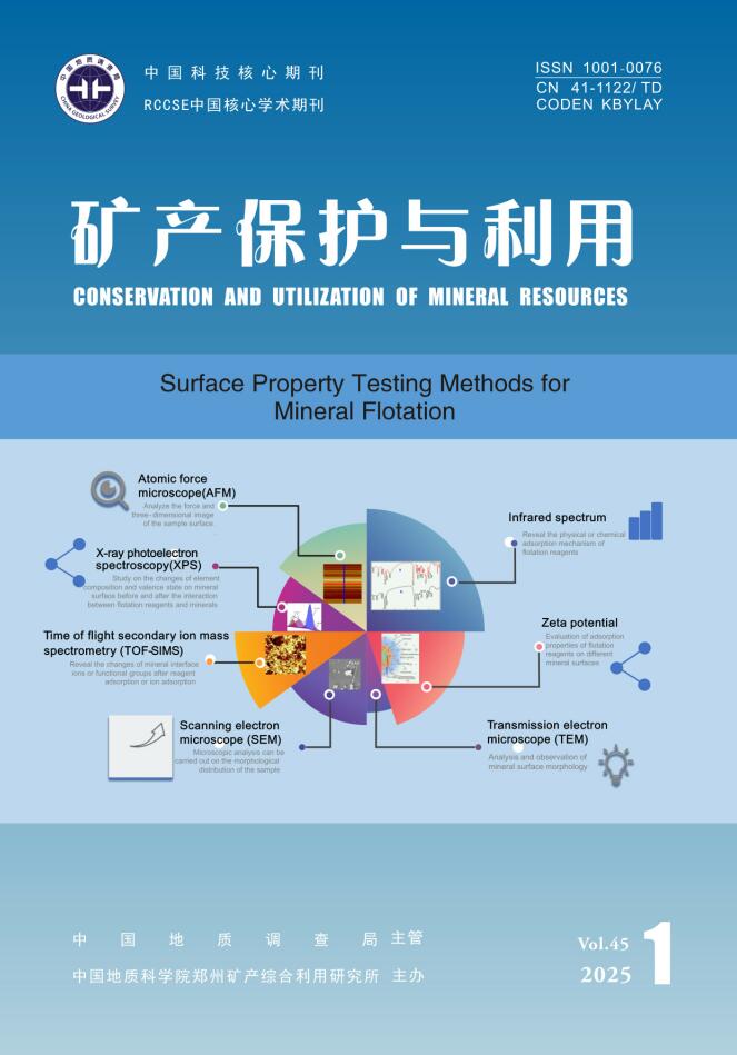
Figure 1.
AFM working principle[2]
-
Figure 2.
AFM 2D plane profile and 3D height image structure image of muscovite mica sample[7]
-
Figure 3.
STEM imaging schematic[13]
-
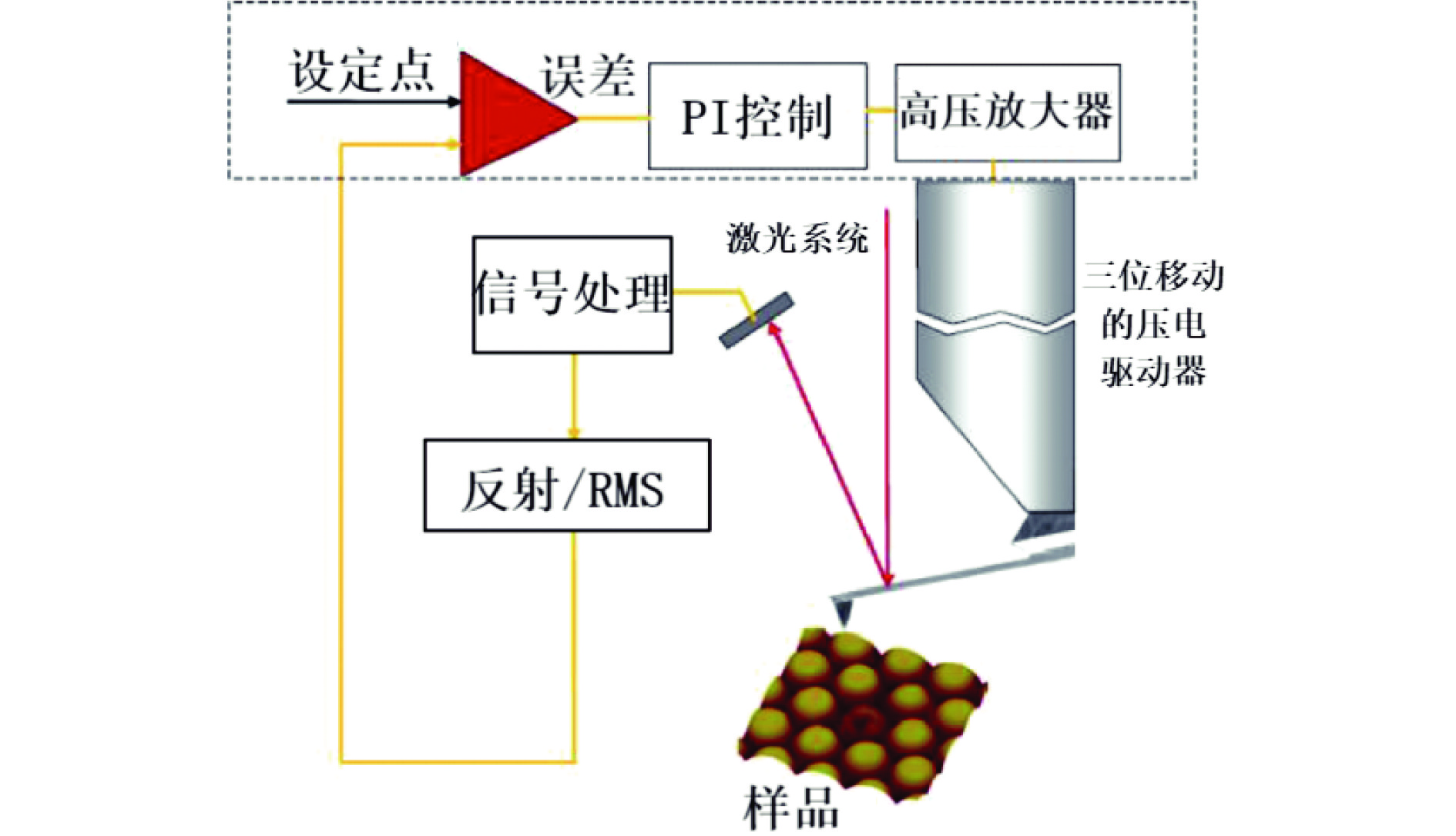
Figure 4.
Bastnaesite (Bst) and novel calcio−cerite form (Syn) form nanoscale bulk derived structures[20]
-
Figure 5.
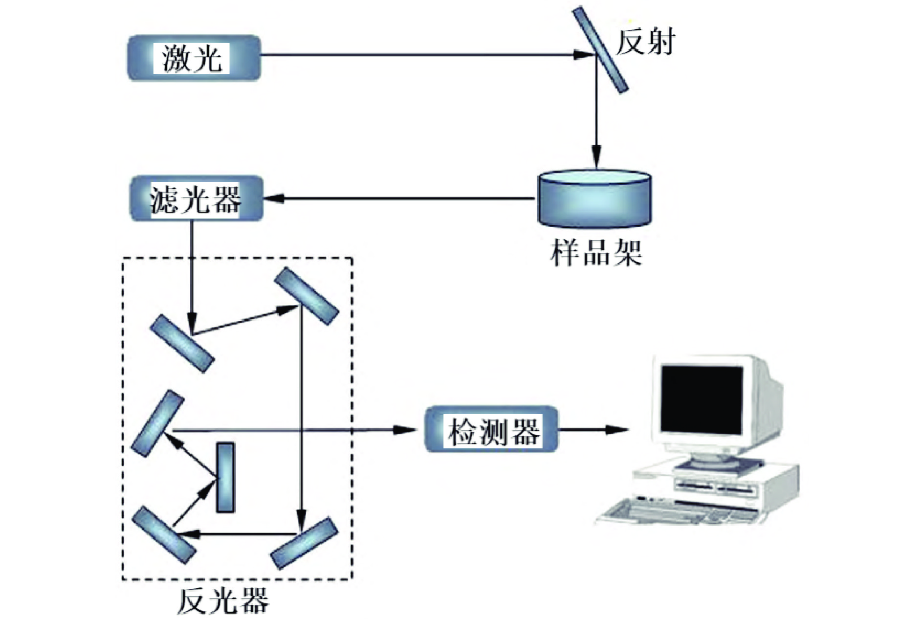
Schematic diagram of scanning electron microscope structure diagram[22]
-
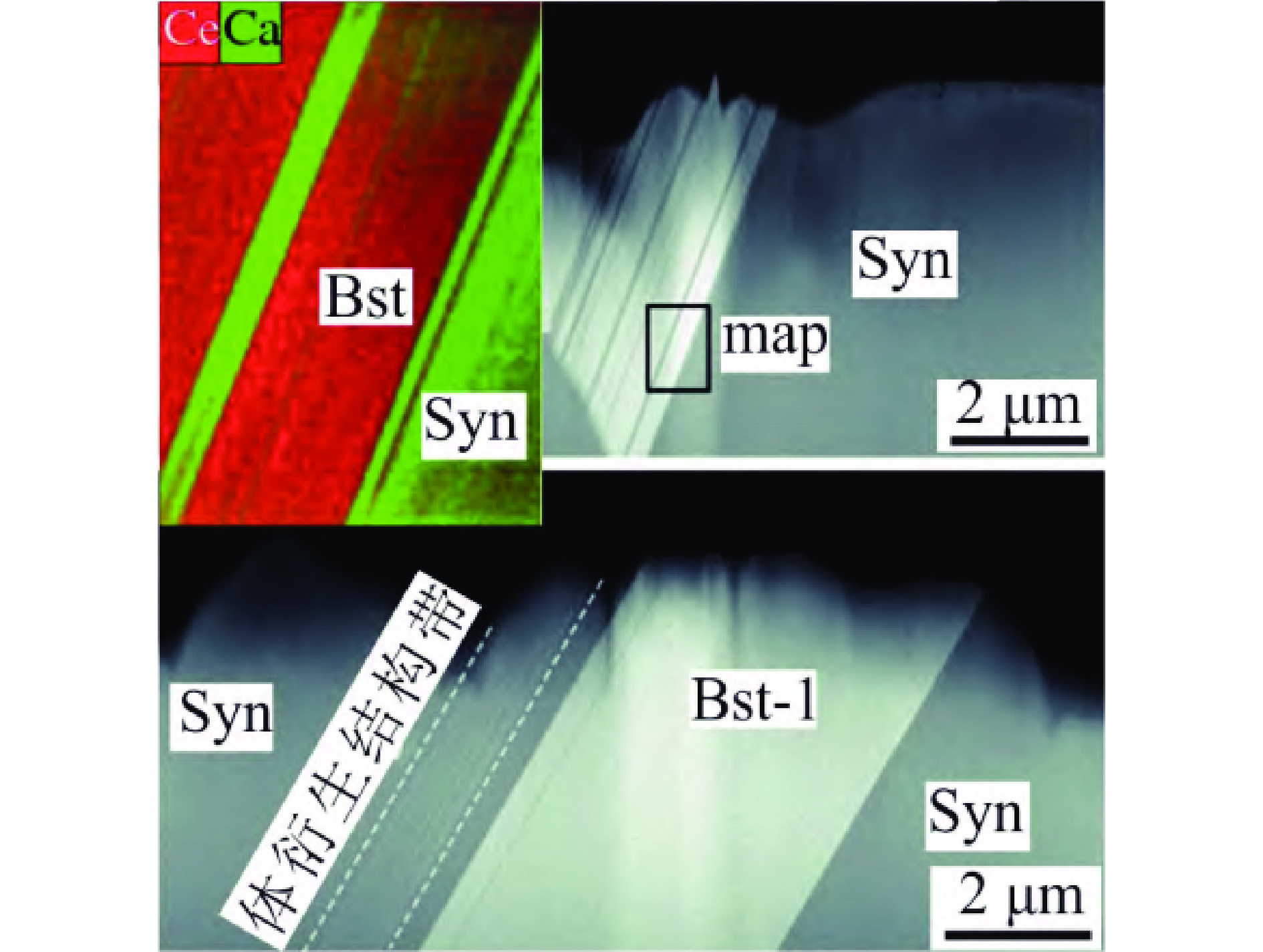
Figure 6.
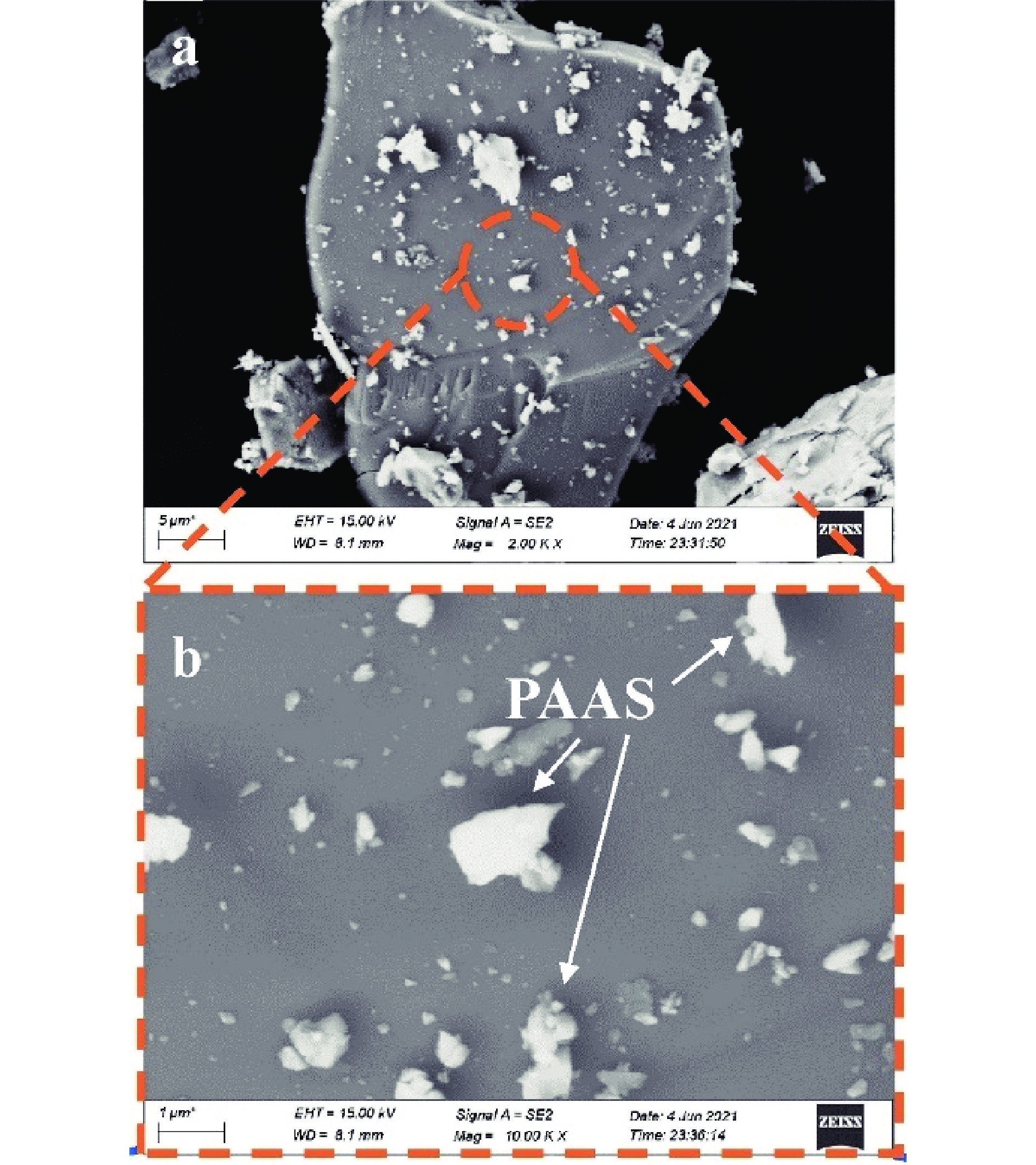
SEM morpHology of hematite surface after PAAS adsorption[24]
-
Figure 7.
SEM images: (a) Malachite treated with Na2S; (b) Malachite treated with (NH4)2SO4+Na2S[27]
-
Figure 8.
(a) Differential ATR−FTIR spectra of pure agent BHA/DDA; (b) Adsorption of ilmenite by BHA/DDA at different pH (concentration: 0.2 mmol/L) [55]
-
Figure 9.
Diagram of Raman spectrum structure[60]
-
Figure 10.
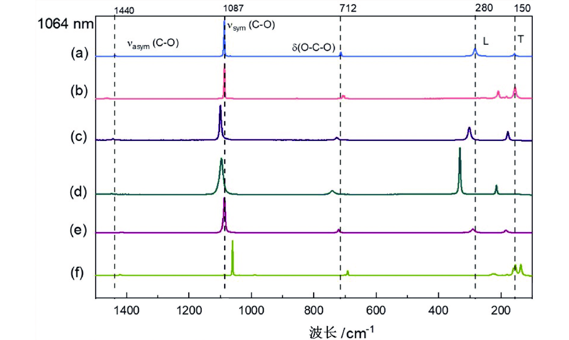
Raman spectrum at 1064nm excitation source of calcite (a), aragonite (b), dolomite (c), magnesite (d), rhombosite (e) and pyroxene (f) [62]
-
Figure 11.
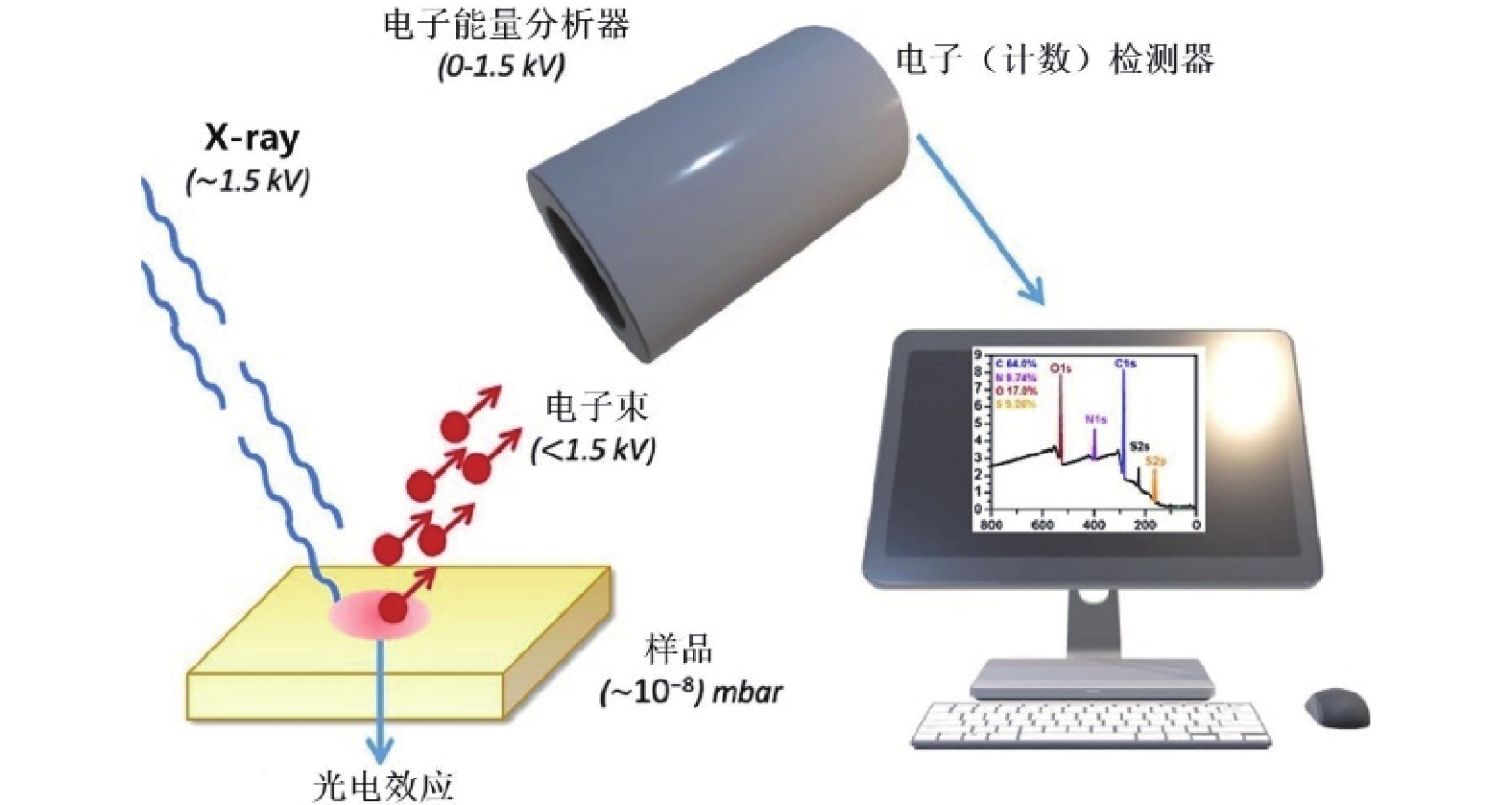
XPS principle diagram[65]
-
Figure 12.
Cu 2p XPS spectrum of malachite treated by Na2S[27]
-
Figure 13.
XPS scanning curves of (a) bare bastnaesite, (b) hydrogenated bastnaesite, (c) OAHD−Ce3+ precipitates, and (d) OAHD[74]
-
Figure 14.
Schematic diagram of TOF−SIMS principle[76]
-
Figure 15.
TOF−SIMS image data of interaction between pyrite surface and EX collector (before and after oxidation modification by Fenton reagent) [84]
-
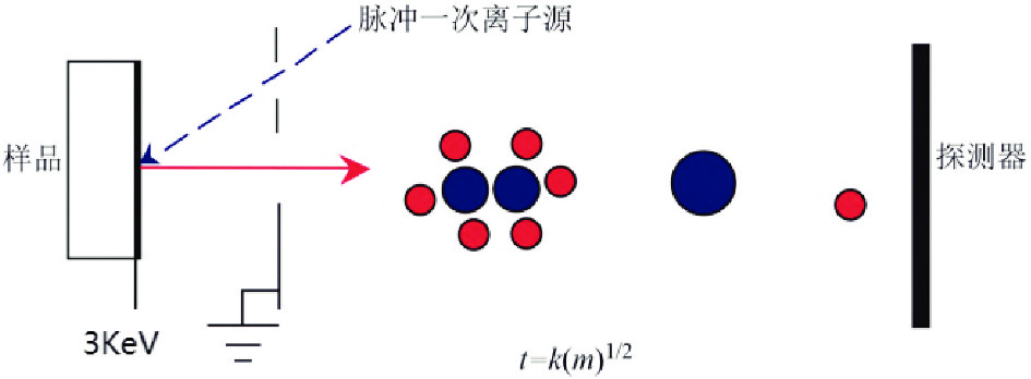
Figure 16.
Image of cationic fragments C2H5N2O+ on the surface of arsenopyrite (before (a) and after (b) adding PASP) [85]